Glaucoma In Your Dog Or Your Cat
Ron Hines DVM PhD
 What Causes Those White Spots On My Cat’s Eyes?
 Corneal Eye Ulcers In Dog & Cat
 Bartonella Related Eye Problems
 Cloudy Pupils In Older Dogs And Cats
When the pressure of the fluid in the front chamber of your dog or cat’s eye rises – it is beginning to develop a glaucoma. Anything that interferes with the normal drainage of aqueous fluid from that anterior portion of your pet’s eye increased intraocular pressure or IOP. Glaucoma is a less commonly identified problem in cats than it is in dogs. Veterinarians do not know if that is because cat develop the problems less frequently, or if cat owners and veterinarians are just less likely to notice their more subtle early warning signs and perform pressure reading on the cat’s eyes. (read here & here)
Unfortunately, glaucoma is a progressive disease. Veterinarians have no cures for it. But when it is detected early, they can often delay the degenerative process with medications. If you want to make every last effort to preserve your pet’s vision for as long as possible, your regular veterinarian will probably refer you to a veterinary ophthalmologist specialist as soon as glaucoma is suspected. But I want you to keep in mind as you read on, that even if glaucoma does take away your pet’s vision, dogs and cats, even with with limited or no vision, remain just as happy and content during their remaining lives. Their other senses and your presence continue to bring them pleasure and contentment.
What Are The Signs Of Glaucoma?
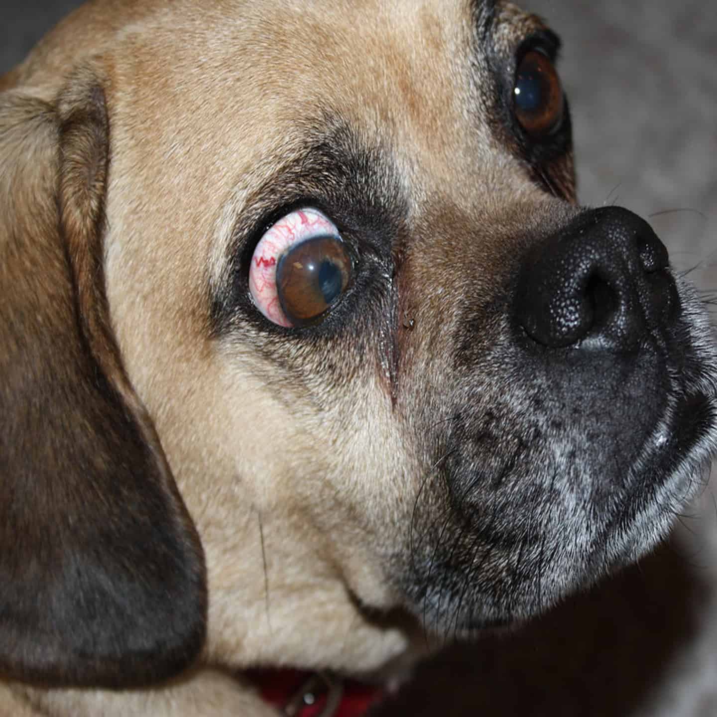
The first sign of glaucoma is often “blood shot” eyes and enlarged pupils which do not constrict normally in bright light or sunshine. The first photo on the top of this page depicts that.
The problem is often first mistaken for conjunctivitis, dust exposure or perhaps an eye allergy or misplace eyelashes. When those pets are given common steroid-containing eye drops or ointments, their eyes often do get better – temporarily. Veterinarians see so many eye irritations of those sorts that steroid-containing eye drops are frequently dispensed.
What Tests Will My Veterinarian Run?
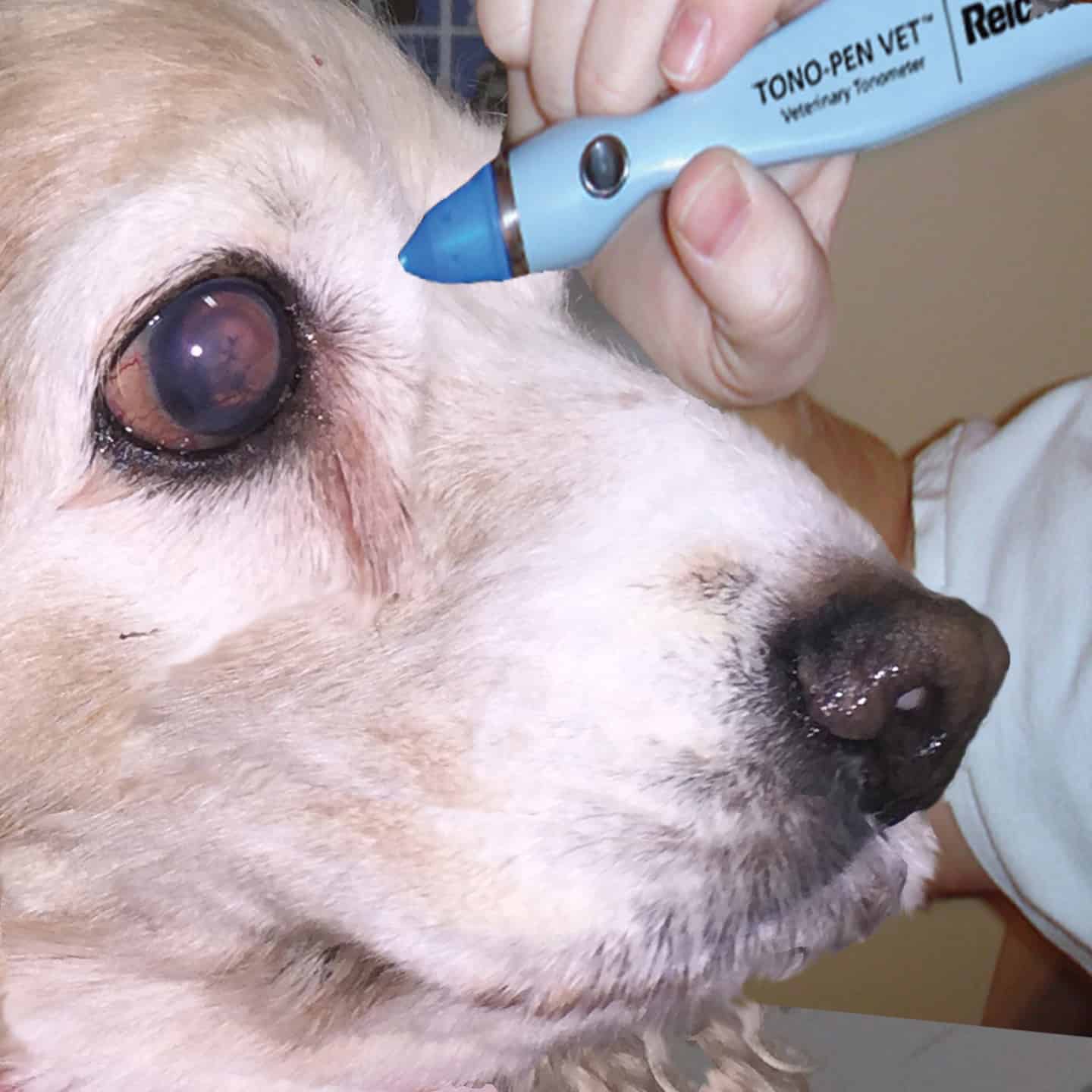
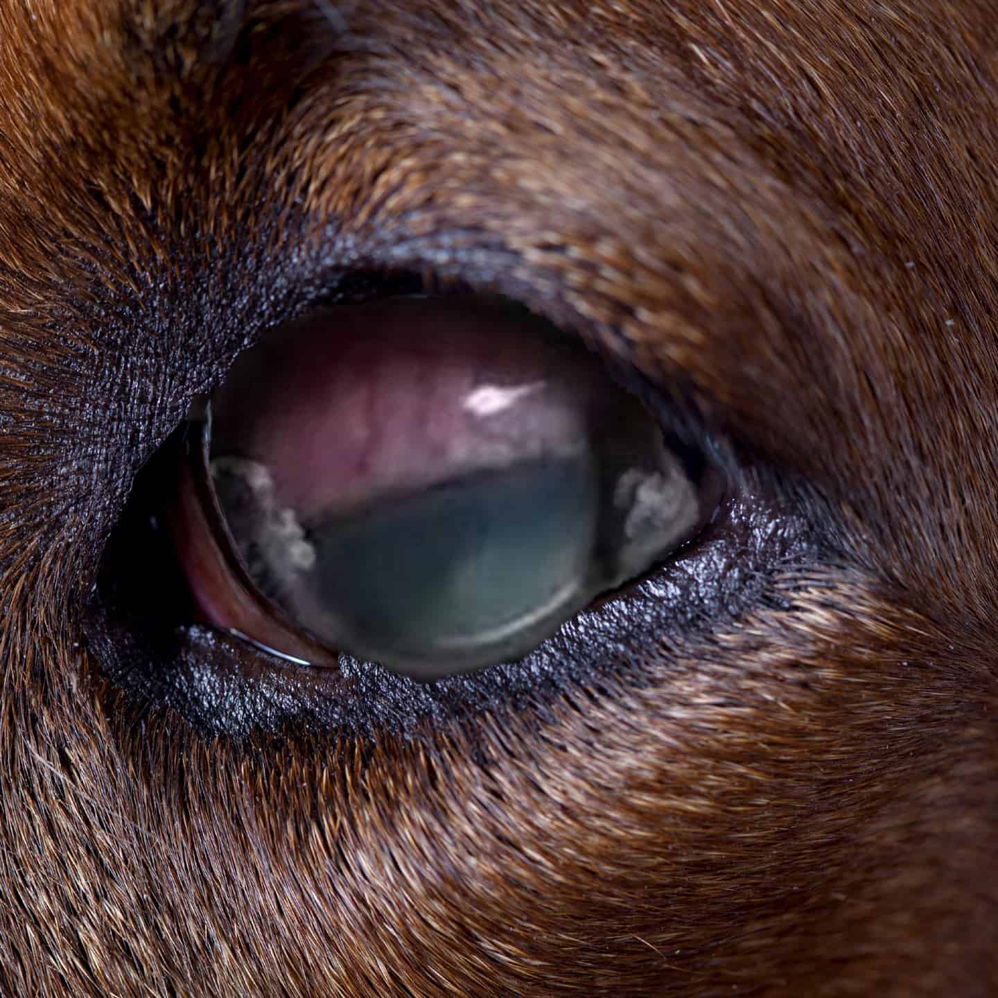
It is always wise to have the intraocular pressure of your dog or cat’s eyes checked (with a tonometer as pictured above) whenever there is even a slight suspicion that the problem might be more than simple eye irritation. This intraocular pressure test is the only way glaucoma can be diagnosed early. (read here) By the time the size of your pet’s eyes actually enlarge, vision has already been permanently damaged. If a developing glaucoma is not treated early, your pet’s eye will also begin to appear cloudy as in the photo above. By this time, most vision is usually already lost and veterinarians have nothing that will restore it. Retinal degeneration, caused by increased pressure within a pet’s eye, can occur very rapidly. (read here) Ophthalmologists have a “30-30 rule”. If the pressure in your pet’s eye stays above 30 mm for thirty hours the pet is likely to lose vision in that eye.
Checking the eye pressure in your pet is not painful. The whole test only takes a minute or two. The older Schiøtz tonometers actually touch your pet’s cornea. So, once a drop of local anesthetic has been placed in your dog or cat’s eye, its head must be held steady to prevent injury. A few dogs and cats might require tranquilization if that apparatus is used. The newer tonometers just puff a small blast of air. All calming medications can affect intraocular pressure. So, they are best avoided whenever possible. (read here and here)
Normal eye pressure (IOP, intraocular pressure) in dogs and cats varies slightly depending on the technique of the person performing the test and the particular device used. But generally, a reading greater than 25 mm (25 mmHg) indicates glaucoma is present. Sustained pressure of 50 mm or greater permanently damage the optic nerve and retina. So, with a reading of 50 or more, your dog or cat’s vision has already been permanently lost – or soon will be. If the test indicates that glaucoma is present, a second test, called gonioscopy will determine if the glaucoma is the wide-angle or narrow angle type.
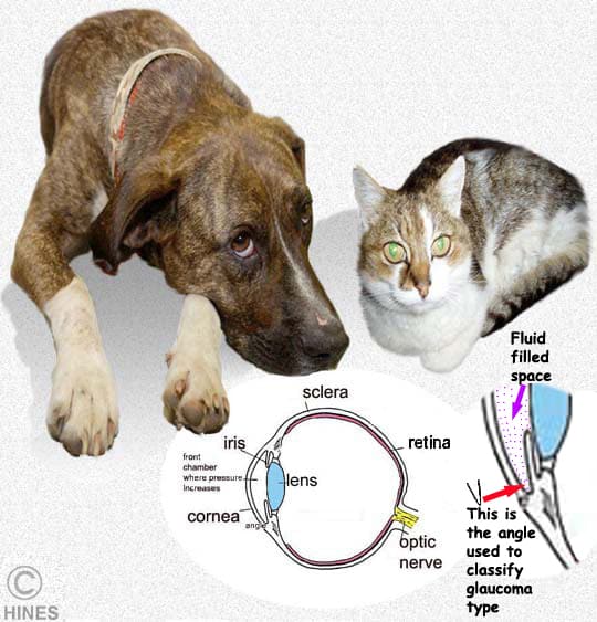
What Are The Different Types Of Glaucomas That Develop In Dogs And Cats?
Ophthalmologists divide glaucomas as being primary or secondary and as to being open angle or closed (narrow) angle. Enlarge the next photo to see the angle involved.
Primary Glaucoma:
Primary glaucoma is a genetic, inherited problem. They are due to abnormal eye anatomy. Veterinarians see these glaucomas most often in cocker spaniels, basset hound eye becomes involved. The results are a complete loss of the pet’s vision. There is no cure. Primary glaucomas are less frequently seen in cats. However, they have occurred in Siamese cats as early as at 8 weeks of age. (read here)
Secondary Glaucoma:
Secondary glaucomas are more common than primary glaucomas that occur early in a pet’s life. Secondary glaucomas form when events take place in the eyes of mature dogs and cats that prevent normal fluid flow and drainage through the trabecular meshwork of the eye. Veterinarians know very little about what causes secondary glaucomas; but cataracts, chronic eye inflammation (uveitis), trauma to the eye, genetics and medications containing corticosteroids have all been discussed. The shape of your dog’s head, driven by its genetics, also influences its susceptibility to a variety of eye issues. (read here) However, what these events that predispose to adult glaucoma might be, why they happened and how we might prevent them is something that veterinarians just do not know. (read here) Glaucoma is also divided into open angle and closed or narrow angle glaucoma. Narrow angle glaucoma is the most common in dogs, although beagles have a high incidence of the open form.
What Happens As Glaucoma Develops?
As I mentioned, when your dog or cat develops the more common, narrow angle type of glaucoma, pressure within its eye often rises quite suddenly. Dogs and cats that develop the much rarer form, open angle glaucoma, develop it slowly over a matter of months. Both forms are quite painful. Your pet might paw at its affected eye or rub its head along the floor. The blood vessels of the white portion of its eyes (its sclera) often stand out prominently and the normally-clear window portion (the cornea) often becomes cloudy or bluish. Bright light might bother your pet. Some dogs and cats lose their appetite and become depressed due to the pain, but most are quite stoic. As this eye problem progresses, the affected eye will appear larger, and its pupil / iris will not contract (constrict) as it should in bright light. By that time, it is too late to save a pet’s vision. This makes glaucoma a medical emergency. In some pets, vision has been permanently lost within less than a day. Please do not feel guilty or feel that your veterinarian could have or should have saved your pet’s vision if you had just arrived earlier. That is very rarely the case.
What Medications Are Available That Might Help My Pet?
Unfortunately, the medical treatment of glaucoma is more successful in preserving vision in humans than it is in dogs and cats. You probably have a friend or relative with glaucoma whose vision has been preserved for many years with topical and oral medications. But dogs and cats usually have a different type of glaucoma than we humans do. And their type, narrow angle glaucoma, does not respond nearly as well to medications.
Your Pet’s Treatment Plan depends on the stage of the problem. The Goals Of Treatment Are:
 Reduce the pressure within your pet’s eye
 Reduce the amount of aqueous fluid that your pet’s eye produces
 Increase the amount of aqueous fluid drainage
 Provide sufficient pain relief for your pet
Medications That Suppress Or Remove Fluid Production Within The Anterior Portion Of The Pet’s Eye:
Osmotic Diuretics
These medications quickly reduce fluid pressure within the eye. That reduces pain and dangerous pressure on the retina and optic nerve. None are appropriate for long-term use, but they are appropriate for emergency situations. Manitol, for example, when given intravenously, will reduce intraocular pressure withing 15 minutes. Oral glycerin is also effective within 30 minutes but is likely to make pets vomit. Although they dehydrate all portions of the eye, they have more effect on the vitreous, shrinking it and allowing better drainage of the more frontal portions of the eye. Those effects last up to 5 hours. Other than in head injuries, they are unlikely to afford long-term benefits to pets with glaucoma.
Fluid Suppressors and Carbonic Anhydrase Inhibitors
These products decrease aqueous production through their effects on the ciliary bodies. They include Acetazolamide (Diamox®), Dichlorphenamide (Daranide®), dorzolamide HCl (Trusopt®), and brinzolamide (Azopt®). Some (e.g. Timolol-dorzolamide) are used in combination with one or more of the other medications. Most CAIs must be applied to the eyes every 8 – 12 hours. In some pets, they can cause eye irritation and an unpleasant taste. Two, methazolamide, and acetazolamide can be given orally. If your pet finds one formulation unpleasant, try another.
Prostaglandin Analogs And Other Medications To Constrict The Pupil For Better Drainage
Prostaglandin analog eye drops sometimes lower intraocular pressure by increasing aqueous humor outflow. They contract the pupil (miosis) and in doing so, open the space (iridocorneal angle) for improved fluid drainage. Two are brimonidine (Alphagan®) and latanoprost (Xalatan®) others are bimatoprost, tafluprost, and travoprost. They perform best for open angle glaucomas. I previously mentioned that narrow angle glaucoma is the most common type affecting dogs. These medications take about 2 weeks to have their maximum effect. The usual dose is one drop once a day. These are human products. If the pet is small, they may need to be professionally diluted. Other miotics perhaps occasionally still dispense are pilocarpine, physostigmine and demacarium bromide
Beta-Blockers
Timolol, is the most commonly used beta-adrenergic receptor blocker used to treat glaucoma. It often helps reduce the amount of fluid present in the anterior portion of your pet’s eye. It is available in eye drop and oral form. Both must be used with caution in dogs with other health issues because the drug also affects the heart and kidneys. The eye drop form can cause irritation.
Is Surgery An Option?
Successful dog and cat surgery to halt glaucoma is in its infancy. Veterinary ophthalmologists use two approaches to the problem – both borrowed from human treatments. The first is to decrease the amount of intraocular fluid produced in the the anterior chamber by your pet’s ciliary bodies. The second is to increase the ability of the fluid to leave the eye through the placement of a shunt – a small implanted tube that drains to the exterior of the eyeball. Until recently, veterinarians attempted to freeze and destroy the fluid-producing ciliary bodies with liquid nitrogen placed on the eye’s external surface. (cycloablation) A second approach was to attempt to destroy the ciliary bodies with a laser. (transscleral cyclophotocoagulation). (read here) Shunts alone had a lower success rate in keeping intraocular pressure down, so most veterinary ophthalmic surgeons combined both the procedures. Results were mixed. The most recent development in veterinary laser glaucoma treatment is the use of the MicroPulse® pulsed laser beam. (read here & here) By firing the laser in very short bursts aimed at the ciliary bodies, heating within the eye is kept to a minimum. That hopefully reduces post treatment inflammation and collateral eye damage. Sometimes, similar treatments are combined with removal of the pet’s lens (phacoemulsification) to allow better exposure of the ciliary areas that need to be destroyed. Claims have been made that after one or more treatments, 50-67% of the dogs maintained an acceptable intraocular pressure when followed for a year. However, many received the topical medications I mentioned earlier as well. Even if your pet remains visionless, its eye(s) might not have to eventually be removed if interocular pressure can be lowered by these surgical procedures. (read here) When no vision remains in your pet’s eye(s), the goal of complex glaucoma surgery is more likely to be to reduce pain and preserve cosmetic eye structure. Preserving the cosmetic structure of a blind eye in a dog or cat is performed to meet the psychological needs of the pet’s owner, not the pet. Less complex, much less expensive removal of the eye (enucleation) also renders your pet pain free.
You are on the Vetspace animal health website
Visiting the products that you see displayed on this website help pay the cost of keeping these articles on the Internet.