
Corneal Ulcers On Your Dogs Or Cat’s Eye
Ron Hines DVM PhD
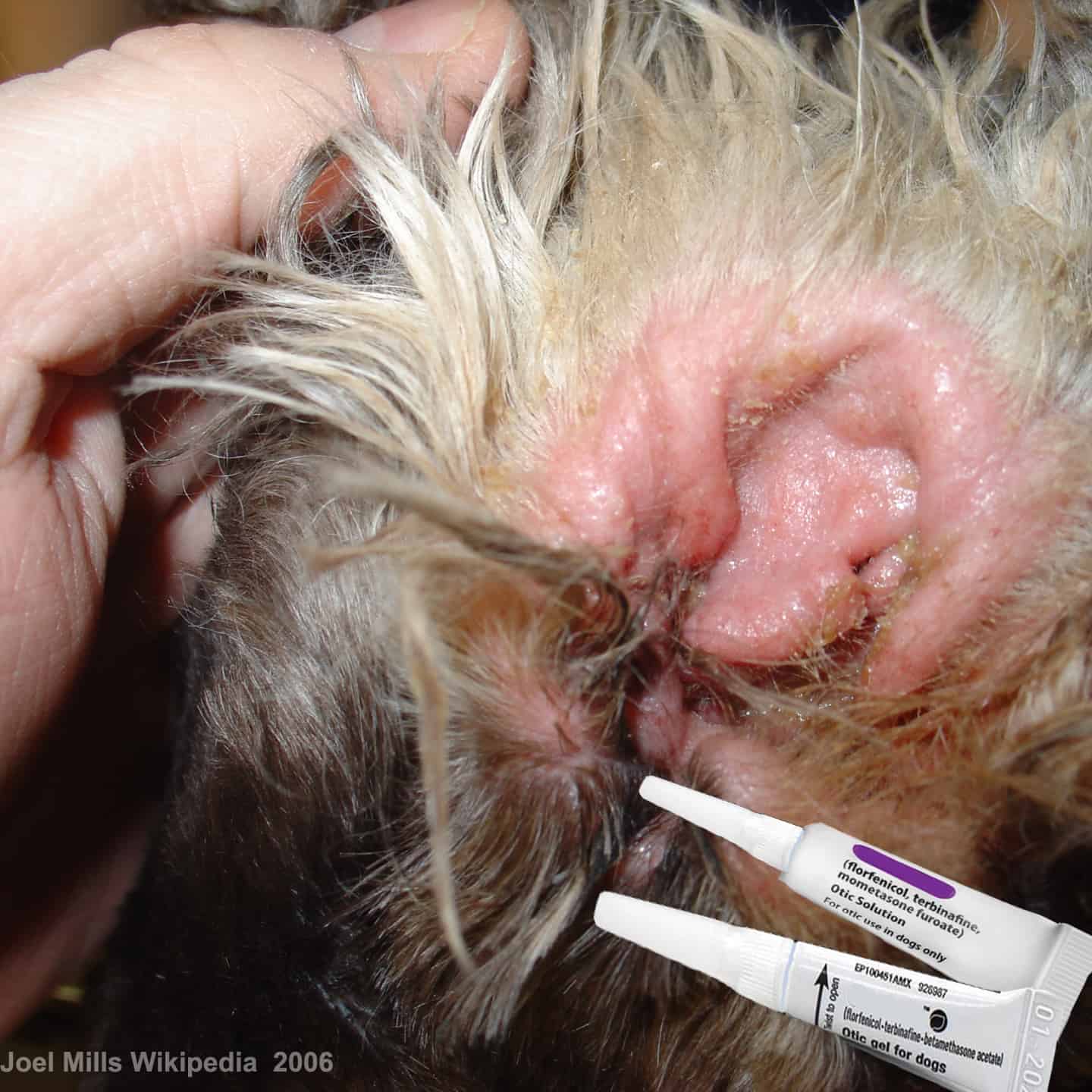 Ear Infections & A Rare Dry Eye Problem?
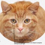 What Causes Those White Spots On My Cat’s Eyes?
The cornea is the clear outer front layer of your dog or cat’s eyes – its window to the world. Corneal cells are the only cells in you and your pet’s body that are transparent. All the other living cells in the body are nourished by tiny capillaries that are about 1/10th the diameter of a human hair. But in order to remain clear, instead of being nourished by blood vesicles that would cause a haze, the cells of the cornea are bathed in a nourishing protective liquid, tears. Because of the cornea’s delicate nature, and its position on the outermost layer of the eye, your pet’s cornea, like yours, is subject to scratches, scrapes and ulceration. That is particularly true in short-nosed dogs who have no protective nose to act as a bumper. When the scrape or scratch is shallow and your pet’s tear production is adequate, your pet’s cornea should heal very rapidly. These sort of shallow scrapes and scratches are most common in young playful pets. Most are so minor that we never notice them. Perhaps a slight squinting for a day that improves with a few drops of artificial tears. But when they are the result of anything from self-pawing, dog and cat fights, the scratch of a thorn, blowing sand, head-out-the-car-window incidents, exposure to infectious bacteria or chemicals, they need professional care. Any sign of corneal milkiness or a closed, or half closed eye is reason enough for an immediate trip to your veterinarian’s office.
Adult dogs and cats are also subject to slower-healing erosions of the cornea we call corneal ulcers. The most common cause of these ulcers in dogs is a deficiency in protective tears due to head conformation (intentionally breeding for bug eyes = exophthalmos) – but corneal ulcers can also occur due to viral, bacterial and fungal infections as well. Adult cats most commonly develop these ulcers due to infection and relapse with the feline herpesvirus-1. In some cases in cats, these corneal defects appear to have an allergic basis, since eosinophils are found in the ulcers in excess (dogs can also be affected with eosinophilic corneal ulcers). (read about eosinophilic complex in cats here) Left untreated, deep corneal ulcers often develop serious complications that destroy or limit vision. You can see the structures involved if you click on the images of the eye at the top of this page.
I mentioned that ulcers of the cornea can be shallow or deep. Once the outermost layer of the cornea has been torn, the area quickly becomes irritated and painful. This causes your pet to squint and its eye to tear. We call that reflex squinting blepharospasm. Corneal ulcers can be quite painful. They begin as an itch – the feeling that something foreign is in its eye. In response, pets will rub the affected eye which just makes matters worse. A discharge usually accumulates in the corner of the eye that is nearest the nose (the medial canthus) and the blood vesicles of the whites of the eye (the sclera) enlarge with blood. If the damaging object is no longer in the eye and the ulcer is not deep, healing is rapid. However, when an infection is present, when the ulcer or cut is deep, when the flow of tears is insufficient or when the pet rubs its eye, the ulcer can progress to affect the deeper structural layers of the cornea. Once the deeper supporting layers of the cornea have been eroded away a bulge can form similar to a sidewall blowout on a tire (a descemetocele). If the descemetocele bursts, the anterior portion of the eye will collapse and the contents will spill out permanently destroying the eye. This is why all corneal ulcers need to be observed closely and frequently by you and your veterinarian.
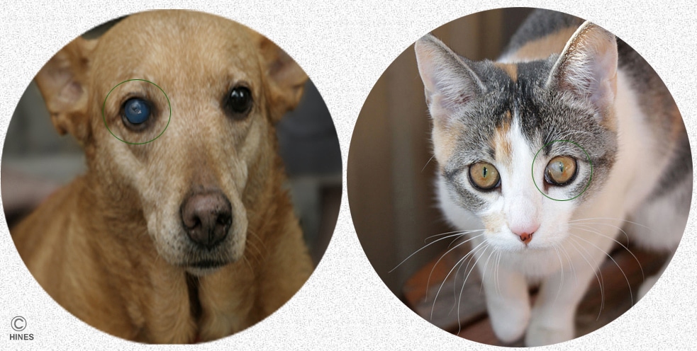
Large, corneal abrasion, and ulcers will cause your dog or cat’s entire cornea to swell with fluid (edema). A normal cornea is ~78% water. When inflamed and edematous, its water content increases. That give the whole normally clear cornea a milky bluish white in color. In an anatomically normal eye, within a few days tiny blood vessels will develop leading to the area of the ulcer in an attempt to furnish nutrients and defensive white blood cells to aid in the healing process. Once the superficial layer of your pet’s cornea has fully healed, those blood vessels should retreat and no more than a small, milky scar should remain.
Corneal ulcers are graded according to their depth. If only the outer layer is lost the lesion is called a superficial corneal ulcer. Those ulcers generally heal without the help of new blood vessels. When more than one half the thickness of the cornea is affected it is called a deep corneal ulcer. As I mentioned earlier, superficial traumatic ulcers in health eyes heal in a matter of days. Deep ulcers can take several weeks to heal. It is the deep ulcers that require new blood vesicles to grow into the area. These are the more dangerous ulcers. An eye with this problem needs intensive care and protection. They can also leave a more substantial corneal scar. When there are underlying causes for these ulcers, some refuse to heal or heal only to re-ulcerate again. Those types of ulcers are considerably more common in pekingeses, pugs and dogs with similar face conformation.
How Will My Veterinarian Diagnose Corneal Ulcers In My Dog Or Cat?
A Fluorescein Dye Test:
Fluorescein dye strips are used to detect and demonstrate that there is a tear or abrasion on the surface of the cornea. First the eye is anesthetized with a drop of topical anesthetic (such as lidocaine or tetracaine). Then the strips are moistened with saline and the dye allowed to flow out over the cornea. The eye is then gently washed with saline to remove all excess dye. In a normal eye, no dye remains on the cornea. Fluorescein dye attaches only to raw abraded areas where it can be seen when viewed with an ultraviolet light source through an ophthalmoscope.
A Schirmer Tear Test:
A second test used to evaluate the eye is the Schirmer tear test. This test measures the quantity of tears present in the eye. One cause of corneal ulcers is a lack of adequate tear flow.
Measure Your Pet’s Intraocular Pressure:
If a veterinarian suspects that the pressure within your dog or cat’s eye is too high (glaucoma) and that this increased pressure and distortion of the shape of the eye is the underlying cause of the ulceration, they usually perform another test. This test uses a device called a tonometer, an instrument that measures pressure within the eye. Ulcers due to the increased intraocular pressure of glaucoma require a different course of treatment. Read about glaucoma in dogs and cats and its treatment here.
More About The Causes Corneal Ulcers In Dogs And Cats:
Corneal Ulcers Due To Accidents
Scraping and puncture wounds of the eye are the most common cause of corneal ulcers in dogs. Cats are less likely to poke their eyes with blunt or sharp objects and more likely to damage their corneas with claws while scratching or fighting. These ulcers are often linear or oblong in shape. If they are not deep, they tend to heal quite rapidly.
Corneal Ulcers Due To Eye Conformation
Dogs and cats with bulging eyes are more susceptible to corneal ulcers. This is because bulging eyes are more likely to be scraped and scratched. Misplaced eyelashes (distichiasis) and eyelids that curl inwardly (entropion) or outwardly (ectropion) can also cause corneal ulcers. Shar-pei and Chow Chows commonly suffer from entropion. Hound breeds are prone to ectropion.
Corneal Ulcers Due To Dry Eyes
The film of tears over protruding eyes often does not reach the center. Dryness of the eye is sufficient to cause ulceration. Other pets are born with deficient tear production. Tear flow over the eye can be increased using cyclosporin ophthalmic drops or corrective surgery. The use of antibiotics of the sulfa class has also been associated with dry eye syndrome. The medical term for dry eye is keratoconjunctivitis sicca (KCS).
Corneal Ulcers In Cats Due To Herpesvirus-1 Infection
The rhinotracheitis aka herpes-1 virus can form a carrier state in the eyes of cats causing periodic corneal ulcers. These ulcers come and go with stress. When inactive, the cornea is left with pinpoint, milky white, rounded scars. When this viral disease is suspected and the signs are severe or persistent, many cats receive topical antiviral drugs such as diluted trifluridine (Viroptic®, trifluorothymidine) and oral famciclovir. Topical antibiotic drops or ointments might be given as well to prevent secondary bacterial infections.
The amino acid, l-lysine, has been found by some to assist many cases of rhinotracheitis conjunctivitis resolve. The thought is that this amino acid reduces the amount of another amino acid, arginine, that is present in the cat’s body. Arginine is thought to be necessary for herpesvirus to reproduce. Many cat owners continue the supplement indefinitely. Lysine can be purchased at health food stores. Pick a brand that is propylene glycol-free. You can read more about this viral infection and the many ways it affects cats here, here, here, here and here.
Corneal Dystrophy
This rare condition is characterized by abnormal deposits within the cornea, which contain calcium and cholesterol. In dogs, it is sometimes associated with hypothyroidism.
Breed-related Problems
Boston Terriers and Boxers have corneas that just do not seem to heal well. When these breeds and others with similar head conformation develop corneal ulcers, some veterinarians believe that topical vitamin E solution hastens healing.
Corneal Ulcers Due To Bacterial Or Fungal Infection
Certain bacteria appear to have an affinity for the surface of the eye causing redness and itching. Self-trauma in these cases can result in corneal ulcers. Those cases need to be placed on antibiotic or antifungal eye drops. Veterinarians are uncertain of the underlying causes. Some associate susceptibility to these infections with dry eye (Keratoconjunctivitis sicca) due to the lack of the antibodies normally found in tears (read here) while others believe it is related to generalized allergies (canine atopy). (read here)
Indolent Ulcers
Indolent means lazy. Indolent ulcers are ulcers that refuse or are slow to heal. These type of ulcers usually occur when a ledge of dead corneal tissue surrounds the ulcer. These ulcers can be encouraged to heal if that dead tissue is surgically scraped away. Other causes of indolent corneal ulcers are persistent dry eyes, infections, or concurrent disease in your pet (such as organ failure) that slow the healing process throughout the body. These long-standing ulcers can also be encouraged to heal when the third eyelid is sutured across the defect in a procedure called tarsorrhapy.
Corneal Ulcers That Progress To A Descemetocele
I mentioned earlier that some corneal ulcers become so deep that the inner corneal lining, Descemet’s membrane, balloons out forming a descemetocele, which is similar to sidewall damage on a tire. If that structure ruptures, fluid from the anterior chamber of the eye leaks out through the cornea and the eye collapses. The iris in these eyes becomes highly sticky and will often stick to or protrude through the defect. If and when these eyes heal, the normal anatomy of the eye does not recover. Distortions of the iris in these eyes often leads to glaucoma and the loss of vision in that eye when the iridocorneal (iris to cornea) angle is changed (see the diagram at the top of this page).
Cats Positive For Bartonella Infection
A high percentage of cats with inflammations of the eye are also positive for bartonella. These cats often get better when they are given antibiotics such as azithromycin, doxycycline or rifampin. Azithromycin is probably the best choice because it also kills other organisms that could be involved in these conditions (mycoplasma). When giving these medications in solid form to cats, you should follow the pill or capsule with water so that the medication does not lodge in the throat. Better yet, give the medicine crushed into a liquid form.
General Treatment Of Corneal Ulcers
The treatment option prescribed for your dog or cat will depend on the severity of the ulcer, how long it has been present and what is suspected to be the underlying cause of the problem. Often a number of treatment options need to be tried before one is found that resolves the problem.
Treatment also focuses on controlling inflammation and pain and preventing additional corneal damage caused by self-trauma. Effective treatment also minimizes residual damage to the cornea by limiting scar formation. Dogs and cats with corneal ulcers are in considerable discomfort and pain. These animals squint or keep their eye tightly shut. They might be sensitive to bright light. Atropine eye drops, which dilate the pupil, usually help relieve the pain. This is the same medicine that ophthalmologists use to dilate your eyes before an eye examination. Topical anesthetic eye drops are required during initial deep eye examinations, but with persistent use they can retard healing.
Antibiotic ointment or drops are frequently dispensed for affected eyes. Corticosteroid-containing medicines are rarely used in the treatment of corneal ulcers because they also retard the healing process. If a pet is pawing at its eye needs to be placed in an Elizabethan collar or have its paws bandaged. Eyes should be rechecked by your veterinarian frequently. Fluorescein dye tests help gauge the healing process.
Deep corneal ulcers require more complicated treatments. These pets often require hospitalization. If Descemet’s membrane is bulging through the ulcer this is really an emergency situation – nothing that can wait for the following morning. Treatment, in those situations, is best performed by a veterinary ophthalmologist. Sometimes reinforcing the area of the ulcer with very fine suture material and then sewed a patch composed of an attached slice (flap) of the third eyelid over the ulcer to shield it and bring it additional blood supply is required. When Descemet’s membrane has burst and the eye has already leaked its contents, the outcome of this surgery is less successful. Some of these eyes cannot be saved and are best removed.
I mentioned earlier that superficial ulcers that are slow to heal (indolent ones) sometimes need to be scraped clean of dead tissue, especially around the margins of the ulcer, in order for new tissue to spread over the defect. This is carefully done under general anesthesia with a small specialized scalpel blade. Tissue adhesives, can be used to provide protection while these ulcers heal.
Ointments and drops need to be applied very frequently to affected eyes. Often every two to four hours when treatment is begun. During this time, you need to observe your pet closely so that it does not rub or scratch the eye.
Pets that develop corneal ulcers because of insufficient tears (keratoconjunctivitis sicca) or a bulging eye are often placed on cyclosporin (aka cyclosporine, ciclosporin) eye drops supplemented with artificial tears. An alternative to this is surgically transplanting the duct of the salivary gland to add additional lubrication.
It is important that your veterinarian check your pet’s eye after 3-4 days of treatment. Most ulcers will have healed in that time, but some will require additional time. If the ulcer has not healed in two weeks, treatment needs to be reevaluated and other procedures such as scraping (debridement) or a corneal patch might be required. If you see the slightest change for the worse, your pet needs to visit your veterinarian immediately.
You are on the Vetspace animal health website
Visiting the products that you see displayed on this website help pay the cost of keeping these articles on the Internet.
