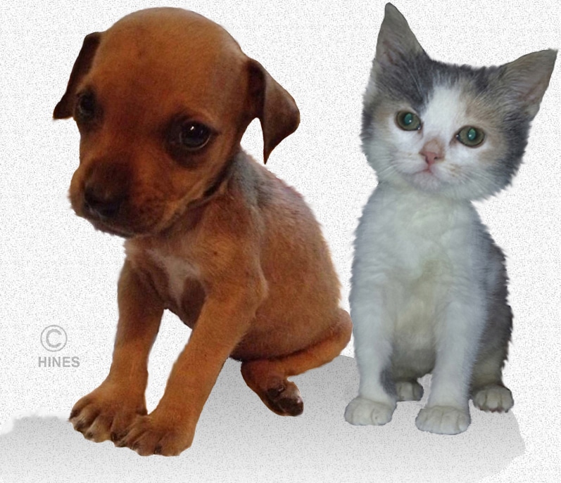
Why Is My Puppy Or Kitten A Runt?
Ron Hines DVM PhD
 What Does My Dog’s Breed Tell Me About Its Health?
There are healthy litters in which all the offspring are a bit small. The larger the litter, the smaller the puppies or kittens tend to be at birth. But that slightly reduced size should affect same-sex individual babies almost equally. Those babies put on weight and grow normally as soon as they are eating on their own when their food supply is adequate and nutritionally balanced. Those large litters also tend to arrive a day or two earlier; which also accounts for the smaller size of each offspring.
There is really no agreement among veterinarians, or anyone else for that matter, as to what constitutes a runt or even how long a dog or cat’s neonatal period (its puppyhood or kittenhood) really lasts. For the purpose of this article, think of a neonate as a puppy or kitten that is less than 7 weeks old and a runt as weighing at least 25% less than its same-sex littermates.
Your kitten or puppy’s neonatal period is one of dramatic growth rate and change. Vital organs are rapidly maturing and taking on their adult responsibilities under the direction of the “anatomic” genes they inherited – the ones whose job it is to guide proper organ development. Through processes called transcription and epigenetics, those crucial genes switch on and then off to guide the developmental process. By about 12 weeks, the job of most of them is complete, and they switch off – for the most part, never to give instructions again.
Even when your puppy or kitten’s development in the womb goes awry (wrong), many youngsters have a normal birth. That is because, to a large extent, puppies and kittens were freeloading on their mother’s metabolic and hormonal systems until the moment they were born. In the womb, it was their mother’s liver, lungs, kidneys and endocrine glands that carried their burden and met their needs. It is only in their neonatal (infant) period that defects in their own metabolism become apparent. This is the most precarious period in a puppy or kittens life – a bird taking wing. And those that are born star-crossed are at the most risk for survival.
It is quite a challenge for me when a newborn kitten or puppy is brought in because of the concerns of its owner. Often these offspring are quite ill by the time they are presented, and they are in a steady downward health spiral. Obtaining blood samples on so small an infant is difficult enough when a baby is healthy. Collecting blood samples can be hard to impossible once an infant is chilled, dehydrated or in other states of circulatory collapse. Just as importantly, many of the standard exams that we veterinarians rely on to direct us to the source of the problem (reflexes, heart and lung sounds, vision, balance, pain sensation, etc.) yield less information in neonates because these systems are not yet fully developed. Another problem is that the signs of a great many different illnesses in puppies and kittens result in very similar signs: subnormal body temperature, difficulty breathing, bluish gums (cyanosis), dehydration, restlessness, diarrhea and crying.
Some veterinarians with specialized training are experts in evaluating the ultrasound images of puppies and kittens while they are still in their mother’s womb. They have mixed success in evaluating pre-birth fetal health. They pay particular attention to the heart rate of each fetus, and the presence of meconium (fetal feces) within the amniotic sac. In human pregnancies, restricted umbilical cord blood flow rather than genetics can also cause slower than normal prenatal growth. That might occur in dogs and cats as well. Veterinarians really don’t know. Doppler velocimetric ultrasound is a common method pediatricians use to detect it.
Puppies and kittens that are markedly smaller at birth are always at a greater risk. That risk is greatest during their first week of life. Which youngsters are at the most risk can also be predicted by measuring their early weight gain (that is, weight gained over the youngster’s first 48 hours). The lower the percentage of weight gain during that critical period compared to their littermates, the less likely the pup or kitten is to survive. That weight gain needs to be at or above 4% (preferably considerably more) and both puppies and kittens should double their weight by 10 days. When that isn’t happening, I tell my clients that the kitten or puppy needs to be pulled and hand-raised. In one canine kennel study I know of, the age of the mother, the size of the litter and the sex of the puppy had no influence on puppy survival. Undersized kittens face the same obstacles.
How Common Are Early Puppy And Kitten Illnesses?
When I worked at the NIH, about 17% of the foxhound puppies born at our Poolesville, MD facility, died before they were completely weaned (before~7 wks). Between weaning and 30 weeks of age, another 4% were lost. (read here) But NIH did not pursue the causes of those deaths in detail. In institutional animals settings as well as at large production breeding kennels (puppy mills), puppy and kitten loss are usually higher than in affectionate family home-born dogs or cats. That is due to stressful communal living, distracted mother dogs, emotionally detached personnel, respiratory infections and a pervasive cost-vs-benefit mentality. An institution or commercial kennel or cattery is never a substitute for a loving home.
There are other less sinister reasons why large non-commercial facilities like the NIH are unlikely to to give individualized care to high-risk offspring. Research facilities need uniform, predictable animals for their research. The less genetic diversity within their animal colonies, the more likely that their scientist exploring the causes and cures for disease will discover them.
Another study performed in Norway was more sophisticated in its attempts to explain the cause of early puppy deaths. But like the NIH, they made no attempts to assist puppies that were falling behind. (read here) In an attentive home environment, with a small number of dogs or cats, infant mortality should not exceed 10 – 12%. Mortality rates in the offspring of random bred cats in well-managed situations is quite low (~ 8-10%). In contrast, feral cat colony kitten mortality is about 75%. (read here)
The number of kittens lost in purebred cat breeding operations can also be quite high – 27.3% in one study, 30% in another. That is not necessarily because of poor cattery conditions. Many trendy purebred cat breeds have too small a gene pool for reliable breed health. That is, they are highly inbred. Inbreeding increases the chances of birth defects, infant mortality and life-long health issues in cats and dogs. (read here & here)
When a breeder’s mortality or the number of undersized offspring are high, that person needs to reexamine his/her breeding program and philosophy and take steps to limit inbreeding and restore healthy genetics – even if that means introducing toms & queens, studs & bitches that are not Show winners or “type” for their breed. In my opinion, that is the only ethical and humane thing to do. I am not the only veterinarian who believes that. (read here)
Puppies that were of normal weight at birth do occasionally die suddenly with no explainable cause. Occasionally, that has a virus cause. (read here) Many pregnant queens carry the herpes virus of cats (cat flu). If that virus is a threat to their newborn kitten or a cause of stillborn kittens is unknown. Similar animal herpes virus do have that ability. (read here)
Do Runts And Small Offspring Occur That Have Nothing To Do With The Youngster’s Genes?
Yes.
There are many causes of smaller-than-normal puppies and kittens that have nothing at all to do with their genetics. Here are the most common ones:
A Puppy or Kitten’s Position in Its Mother’s Womb = Intrauterine Position
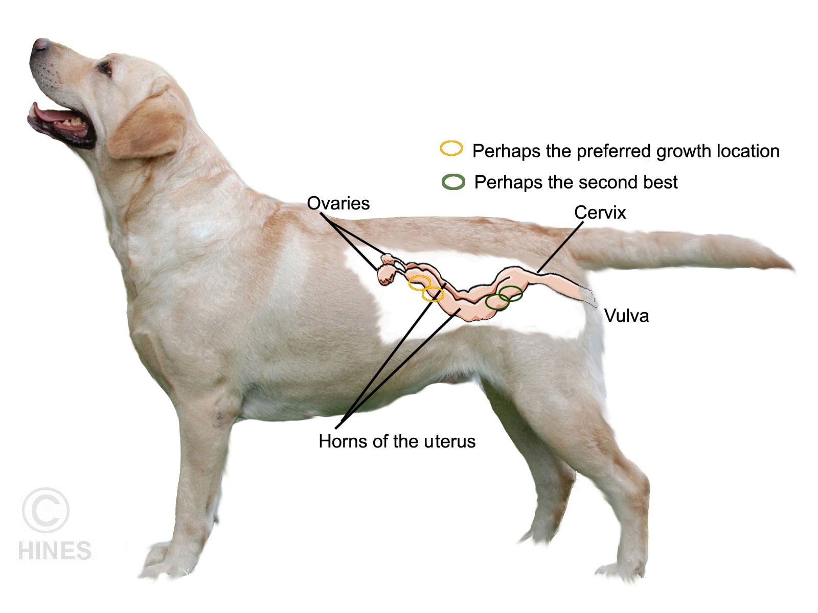
As I mentioned earlier, sonograms and x-rays are very good at discovering undersized kittens and puppies while they are still in their mother’s uterine horns. However, veterinarians cannot accurately tell from those images where the infant is located in the mother’s uterus. There are probably ways that could be done, but I do not know of anyone who has described it. All animals that give birth to multiple offspring (that have litters) are called multiparous animals. They all have similar reproductive anatomy: a small uterus with long horns in which the babies develop that lead up to each of its two ovaries. Conditions within the first third, second third and last third of the uterine horns can be quite different due to differences in how the mother’s blood nutrients, oxygen and blood-carried hormones circulate in each area. Embryos attach to the walls of the uterine horns like passengers lined up in airplane seats plugged into their headphones. Our love for our dogs and cats have spared them from a lot of curious dissection and experiments; but in another multiparous animal, the pig, infant growth is greatest in the first and second seats (positions) closest to the ovary, the slowest growth is near the middle of the uterine horn and better again nearest to the cervix. The piglets that occupy the preferred positions are born larger and the little ones most likely came from the middle. The same thing occurs in rabbits and mice, so I will venture that perhaps similar things occur in our dogs and cats as well – although I have nothing to show you to prove it.
There are other things that are not always uniform in the womb while a kitten or puppy is developing. Male embryos naturally grow larger than female embryos due to the larger amount of testosterone being produced in their bodies. Male embryos actually exude that hormone into their surroundings. That leaked testosterone is known to increase the size of female embryos that are next to them, so they grow a bit larger too. That extra testosterone might even affect that female puppy’s or kitten’s later reproductive potential – its reproductive tract anatomy and fertility.
A male puppy embryo between two females in its mother’s womb will likely be a bit smaller than males next to males. Many years ago, one of my veterinary school professors suggested that as an explanation for small birth weight puppies and kittens. Like the effects of position in the womb, no one I know of has studied this hormonal effect in dogs or cats, but it has been confirmed in rodents, pigs and rabbits. There are probably other potential undescribed variations and defects in a mother’s uterine horns that might not allow a fetus to grow to its intrinsic (natural) potential that are still unknown. (Human multiple-birth pregnancies are different because normal implantation in us is in the uterine body, not the uterine horns – but even there, various environmental factors influence multiple birth baby size.

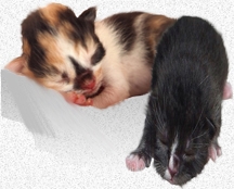
When a fetus is faced with those situations, it is often born with a head disproportionately large for its body – similar to the ones in the image above. It is called brain sparing and some theorize that when nutrients, or oxygen are scarce, it occurs in all mammals. Not only must a mother’s blood deliver nutrients, oxygen and hormones to the developing embryos; it must also cleanse the infant of the waste products of the infant’s metabolism (toxins). So, when blood flow is lessened for any reason, its lessened ability to rid itself of waste products can play a part in stunting as well. None of these points have been studied in dogs and cats. But many studies have examined the phenomenon in humans and animal models.
Once a puppy or kitten is born, those small differences in size can get magnified as the larger youngsters compete better than the smaller ones for milk. That is why it can be best to bottle-raise those smaller offspring after they have had a 24-hour, supervised, feeding on their mother (to obtain colostrum). It is not uncommon for bottle-fed infants to grow at a slightly slower rate than mother-fed infants. We do not know if that is due to the residual effects of whatever made them small in the first place or if there are deficiencies in commercial milk formulas. Over-feeding as a substitute for frequent moderate feedings can lead to diarrhea, bloat, stomach and intestinal stasis, slower growth rate – or worse (In my experience, many more problems occur in kittens and puppies due to too infrequently feedings with too much formula at one time than to underfeeding them).
A Mother’s Neglect Or Rejection

Some dogs and cats are just not cut out to be good mothers. The reasons can be their personality, general health, insufficient mothering hormones (oxytocin, prolactin, endorphins etc.) or just a mother’s immaturity (e.g. her first pregnancy). When these sort of problems occur, they should affect her attitude toward all the offspring in the litter equally.
Some cats and dogs will occasionally reject one or two offspring from their litters. Those are almost always the ones destined to be runts. I do not know anyone who has studied this scientifically, but it’s my opinion that it is primarily low body temperature (hypothermia = less than 95 F/ 35 C for a newborn or notably colder than its siblings). That causes these mothers to lose interest in those offspring. Hypothermia is not a disease in itself; it is the end result of most everything that can go wrong in a puppy or kitten (dehydration, infections, respiratory and circulatory problems, infections, low blood sugar [hypoglycemia], etc.) Placing the offspring in an incubator and bottle-feeding it are the only ways I know to surmount the problem. However, the chances that you will be confronted down the road with health issues in those infants are considerable. A mother’s sense of smell does allow her to identify each of her offspring.
Thermal Issues – Hypothermia Is Hardest On The Smallest In The Litter
Puppies and kittens are quite sensitive to chilling. That is because of their much larger surface skin area in relation to their body weight, and the fact that they cannot shiver to raise their body temperature for their first week or so. They also lack insulating fat and can have problems redirecting blood to their internal body organs (surface–vasoconstriction). These problems are even greater in runts because of their smaller size and greater surface area in relation to their weight. It is not until puppies and kittens are 7–8 weeks old that they can regulate their body temperature efficiently. When an infant’s body is below its normal temperature, a host of bad things occur. It cannot absorb nutrients well from its intestines; it is subject to stomach and intestinal bloat (ileus); its heart does not pump blood efficiently (CRT time increased ); its liver cannot produce blood sugar effectively and its immune system looses its ability to prevent infections. One must never attempt to feed these infants until their body temperature has been restored to normal. Its first few meals should be no more than Pedialyte™, the next few a puppy/kitten formula fed quite dilute. Weakness makes milk aspiration into the lungs considerably more likely. Infants that are already fading are best given their fluids subcutaneously. Warmth is critical – but I have seen more of these little guys pass away due to overheating from the use of human heating pads and heat lamps then I have seen them benefit from them. Hot water bottles are much safer, as are commercial rigid heating pads designed specifically for pets. As I mentioned, chilled offspring in their first few days of life are also considerably more susceptible to infections. Antibiotics like amoxicillin/clavulanic acid can be helpful in preventing that, but they can also contribute to bloat problems. So combining those medications with probiotics might be wise.
Hypoglycemia – Low Blood Sugar Level
Smaller than normal puppies and kittens are more susceptible to low blood sugar levels than their larger littermates. When it occurs in runts, it is almost always together with hypothermia. All puppies and kittens are born with very little fat reserves. They must provide their energy through internal glucose production (gluconeogenesis) and the smallest puppies in the litter have the least glucose reserves. They are the ones that need milk the most; but they are usually the ones that obtain it the least because they are the least able to compete for a nursing position on their mother – particularly during their first 24 hours after birth. From then on, it can be a downward spiral if they do not receive supplemental feeding or your assistance in getting a nipple to feed on. It is also critical that supplemental feeding be extremely small in volume and frequent (every hour) as these infants are at high risk of aspiration pneumonia when more than a drop or two at a time is fed. Table sugar solutions are a very poor substitute (a balanced puppy/kitten formula is much better). Table sugar is sucrose – only half the molecule is glucose (aka dextrose) that the baby needs. Table sugar digestion relies on the enzyme, sucrase, which is normally present in the intestine. But this enzyme’s ability to split table sugar into glucose and fructose is temperature dependent and the babies that need the sugar are generally too cold. The result is fermentation that causes bloat – often severe enough to interfere with their breathing. Hypoglycemia is more common in toy breeds. These are also the breeds most frequently affected by hepatic microdysplasia that you can read about farther down in this article and in greater detail through the former link. Any form of liver disease increases the risk of hypoglycemia.
Dehydration
The bodies of kittens and puppies normally contain more body water than adults. Runts are at particular risk of dehydration because their larger surface area ratio to their weight increases water loss through their skin. Any diarrhea or vomiting increases the problem. So does excessive heat. They are also less successful in replacing their body moisture through suckling. During development in their mother’s body, they relied on her kidneys to maintain a proper fluid balance. After birth, their kidneys have not fully developed the ability to conserve water by producing more concentrated urine or increase the excretion of salt to keep blood sodium levels from rising too high. So, any time a youngster becomes dehydrated, its blood sodium level can go far above normal too (hypernatremia). The signs of dehydration/hypernatremia in puppies and kittens are a doughy skin that does not spring back when it is pinched as well as lethargy. Like just about everything else that affects the newborn, their body temperature drops as well. Mild dehydration can be treated with oral fluids. But severe dehydration needs subcutaneous and/or intraperitoneal injections of balanced sterile fluids (e.g. PSS, D5W).
Lack Of The Mother’s First Milk – Colostrum
The immune system of newly born puppies and kittens is not experienced enough or developed sufficiently enough to produce adequate antibodies of its own. For that, it relies on the antibodies of its mother that are only able to enter its body intact through its intestines during its first day or so of life. The crucial time for this to happen in puppies is their first 4–8 hours after birth. By 16–24 hours, the infant’s intestine is practically closed to the passage of antibodies.
This mother-to-infant transfer is called passive immunity, and it differs from the active immunity that will eventually be produced by the offspring’s own immune system. The mother’s colostrum not only contains antibodies, it contains immune system cells from its mother that produce antibodies and defend the intestines as well. (read here) So thawed frozen colostrum is not as protective as fresh colostrum flowing from the mother. Runts obtain less colostrum. That makes them more susceptible to the infections and septicemias that I mention later.
Other puppies and kittens are occasionally born with defects in their own immune system. Those neonates don’t start to fall behind in growth and general health until their mother’s colostral immunity wears off at about 12–14 weeks – earlier if the mother’s immunity profile is low, occasionally a bit longer if the mother’s immunity was high. Once the mother’s immunity does wear off, that occasional puppy or kitten with genetic defects that prevent its immune system from functioning is in trouble. That will be the time that repeated illnesses begin. (read here & here) Those sort of defects are considerably less common in kittens than in puppies.
Veterinarians have techniques that we thought would supply these important antibodies when the mother was unable to do so. We might have recommended that during its first 24 hours, the infant receives an oral supplement of antibodies derived from a dog or cat’s blood (hyperimmune serum). But the latest study I know of did not find that to be effective. The jury is still in deliberation on that.
Occasionally, it is not a lack of colostrum, but what the colostrum contains that is the problem. Some lactating mothers are more susceptible to breast infections than others. The organisms producing those infections will pass with the milk to the babies. The fact that the mother herself was susceptible indicates that she lacked sufficient antibodies to protect herself. So, the offspring lack them as well.
This problem is sometimes called “toxic milk syndrome”. It can occur even if there is no obvious evidence of a mammary gland infection in the mother. These sort of dangerous bacteria have other ways to move from mother to infant as well (Although I am suspicious that problems would not have occurred had there not been husbandry or underlying immunity transfer problems in the dame).
Delivery Problems
Almost 80% of the time, after one puppy (or kitten) is squeezed out of one of the uterine horns, the next offspring will be expelled from the mother’s other uterine horn. (read here) For a variety of reasons, the birth of one puppy can be delayed. Sometimes the mother is exhausted by the continuing effort to birth a large litter. Sometimes her blood calcium level may not be optimal. Other times, pain, anxiety or distraction make a birth take longer. In those cases, puppies and kittens have been without oxygen (hypoxic) for too long a time and the acidity of their bodies is higher than it should be because they have been deprived of oxygen. Umbilical cord kinks, twists and compression or early placental detachment can cause the same situation. Some believe that puppies that exit tail end first (breach) are also more difficult for the mother to pass. Others have noted that interrupted or slow deliveries (uterine inertia) are more common in mothers that were relatively old when bred for the first time. The interplay of blood calcium level and oxytocin might also be a cause of one or more slow pup deliveries.
Puppies and kittens that have experienced that sort of birth trauma are more likely to be weak (or die in their first 48 hrs) and fall behind in their growth.
Other Maternal Factors
Hypothyroid Mothers
Early embryo development is dependent on thyroid hormones provided by the mother. As the embryos get older, they begin to produce thyroid hormone on their own. We now know some of the genetic errors that make adult dogs more susceptible to hypothyroidism. Although a lack of thyroid hormone early in pregnancy will negatively affect the puppies, I believe that most or all of those hypothyroid adult females are quite unlikely to become pregnant in the first place. When the pup or kitten itself is hypothyroid, it will stay small (I write about that farther down this page). But should a female dog that is hypothyroid or borderline hypothyroid actually produce pups, the signs in the puppies or kittens would probably be similar to those in humans with the same problem; if they go full term, the offspring are actually larger than normal.
The Mother’s Nutritional Status

Nutritionally deprived mothers and those with heavy intestinal parasite loads (or fleas or ticks) tend to produce smaller offspring. But other than the effects of uterine horn position that I discussed earlier, the whole litter, not just one, will be small. Diets do not only need to satisfy the animal’s appetite, they need to be balanced in their protein, amino acid, mineral and vitamin content as well to produce healthy offspring.
Mismatched Blood Types – aka Neonatal Isoerythrolysis, NI, FNI
This is a problem that puppies rarely, if ever, face. It is also a problem that the owners of a kitten(s) from an accidental indoor-outdoor mating are unlikely to see either. But it can be a challenge to purebred cat breeders who have not blood-typed their breeding cats. The story generally goes like this: The queen gives birth to what appear to be normal kittens, and they begin to nurse properly. But some or all of them soon lose their desire to nurse. They become weak with little or no energy. They may die in as little as 24 hours after these symptoms begin. Some pass reddish brown urine and develop a yellowish skin tint (jaundice). Their gums are quite pale. Sometimes only one or two kittens in the litter are affected. Sometimes all are. Also, the degree of illness varies greatly –perhaps depending on how much of the first milk (colostrum) they consumed. Occasionally, the only thing noticeable in a survivor is the loss of the tip of the tail. In the rare puppy that might develop neonatal isoerythrolysis, most symptoms would be the same.
The roots of this problem are antigens – molecules that coat the surface of all red blood cells. These proteins determine blood type. In cats, there are three major blood types, A, B and the rarely encountered AB. Most random bred domestic cats throughout the world have two A genes for the blood type. Most that have AB only express A, so they are A blood type as well (some dispute this). But cats that have BB, are blood type B.
A is the most common blood type among cats in general. But there are some B domestic short-hair cats as well. Their number seems to vary depending on the area of the world in which they live. For instance, the B blood group is said to be more common in India, France, Italy and Australia than it is in North America. However, certain breeds have high numbers of B blood group. Exotics (a short hair version of the Persian), British Shorthair, Turkish Angora, Vans, Cornish and Devon Rex have a high incidence of type B. Breeds that are typically type A are Siamese, Burmese, Russian Blue, Ocicat and Oriental Shorthair. Ragdolls have the highest known prevalence of type AB. (read here)
All A and B cats naturally possess antibodies (alloantibodies) against the blood group they do not possess. Those antibodies begin to appear at about 3 months of age. No one is certain why. Those antibodies will quickly destroy (erythrolysis=hemolysis) red blood cells that do not match the cat’s blood type (in humans but not in cats or dogs, these alloantibodies can pass through the placenta and into the fetus before birth. Dogs and cats have a different, less intimate, type of placenta than humans. So, in them, it does not happen).
Type B mother cats all possess antibodies against type A. If a type B mother cat is mated to an A or AB tom cat, any kittens born with an A blood type can be severely harmed when they drink the mother’s first milk (colostrum) which is high in her anti-A antibodies (how AB kittens will be affected is debatable). Depending on the amount of antibody in the mother’s milk and how much the kitten drank, the blood in the kitten’s body will be quickly destroyed. By 12–24 hours after delivery, however, those antibodies are typically no longer present in sufficient quantity in the mother’s milk to cause problems and are thought to have more difficulty entering the youngster’s blood stream even when they are. (read here) Kittens that are only moderately affected are often slow to gain weight. As I mentioned earlier, a few loose the tips of their tails (tail-tip necrosis).
Medications, blood transfusions and general intensive support of seriously sick infants with NI is rarely successful. They just go too quickly. Some vets attempt to give them intraperitoneal or intraosseous (into the bone) injections of washed type B red blood cells in an effort to replace the A Type cells that were lost. But as early as day three, the A-Type kittens can be producing anti-B antibodies of their own.
So preventing this problem is a much more successful option. It requires blood typing all breeding cats in the cattery and never mating a B-type female to an A or AB-type tomcat. If you insist on doing so, the kittens need to be foster raised by an A mother, for the first 24–48 hours of life. Some breeders just deny the kittens nursing privileges on their birth mother for 24 hours and bottle-feed formula. I do not recommend doing that because colostrum has important health-protective functions.
There are other rare inherited genetic defects that, at first glance, might be mistaken for neonatal isoerythrolysis (although they generally occur considerably later in life than NI). (read here, here & here)
Runts With Defective Gene Issues = The Youngster’s Genetics Are Causing The Problem 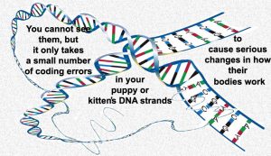
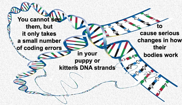
The kitten and puppy problems I discussed up until now often improve as time goes by or when the specific mother-related problem is corrected through bottle-feeding or other means. The rest of the problems that can cause a runt are defects that do not heal themselves. Some of these health issues veterinarians can cure, some they can help significantly, some slightly and some not at all.
I cannot tell you about all of them. First, because I do not have the knowledge nor the time, and secondly because many of the causes have not yet been discovered. But at the Faculty of Veterinary Science of the University of Sidney, Professor Frank Nicholas and his associates make a sterling attempt to do so. Their OMIA website: http://omia.angis.org.au/home/
Unraveling the cause of a genetically based disease is a time-consuming, expensive process. One that few, if any, individual pet owners could financially underwrite. So, we veterinarians understand considerably fewer of these mechanisms than your family physician or pediatrician does. Much of what we do know; we know because scientists studying the problems were financed and motivated to find explanations and cures for a similar disease that occurs in human infants. Dogs and humans share some 350 heritable disorders with us – cats and humans share somewhat less. Neogen is a company with considerable expertise in looking at the genetic makeup (genomics) of living things. Their GeneSeek division offers (or plans to offer) genetic probes (“chip platforms”) that search for genetic defects in pets. Currently, they are beginning to market that service to pet owners through Mars Inc. as the “Wisdom Panel”. But Neogen told me that they can be employed directly to seek out the genetic cause of almost any inherited pet abnormality. (From my conversations with them in early 2016, it appears that their business plan is in its infancy and subject to change.) In 2016, a company affiliated with Cornell Veterinary School announced “Embark”, a similar service, was still in the planning stage.
Are Purebred Pets More Susceptible To Genetic Problems Than Mixed-breed Pets?
Yes.
When you are dealing with purebred dogs and cats, I believe that genetic problems underlie the majority of babies we see with stunted growth. When the infant is the product of a cross or feral mating, I believe that non-genetic, intrauterine, nurturing and psychological issues in the mother outnumber genetic causes. Others do not agree. But most of the genetic diseases pet owners and veterinarians see only occur when both of the pair of genes that control a particular process are defective (a recessive trait). For many genetic problems, the chances of that happening are much greater in dogs and cats that are too closely related – the closer the relation, the greater the risk. For a few, they are not because the tendency is a dominant trait. (read here)
I cannot prove that because no one has examined the issue thoroughly and few intentionally breed mix-breed dogs or cats. A few years ago, I would have just been able to list some of these genetic defects that produce runts and deformed offspring. But over the last 10 years, veterinarians have learned much more as to the specific defective gene(s) that cause these problems. That explosion of knowledge began in the 1990, when Congress, through the NIH, increased funding for the human genome project – a project to name and map each gene pair within us and describe its function. At the forefront of this project is the Broad Institute in Boston (rhymes with road). In 2005, Broad finished mapping the genome of the dog. (read here) In 2007, they published the preliminary genomic map of the domestic cat. (read here) The US government justified adding the dog and cat genome not for us veterinarians and pet lovers; they did it because humans, dogs and cats have more genetic diseases than any other animal species and understanding and attempting to repair or prevent these genetic issues in dogs and cats could have applications in human medicine as well.
In genetics there are as many opportunities for things to go wrong as go right. Most genetic pet problems that veterinarians encounter require that both the dam and the stud carry a defective gene (a recessive carrier); although a few require only one defective gene (a dominant trait). (read here) Some cases of kidney inflammation and portosystemic shunts are dominant traits that require only one defective gene. (read here) Other birth defects require defective genes at multiple locations (polygenetic).
I’ll try to tell you about the more common ones that are based on faulty genes, and some less common ones where the gene(s) responsible have been discovered. There are far too many for me to list all of them. Not all kitten/puppy birth defects are due to gene defects, but 60-70% probably are. Lumped together with the ones whose cause can’t be determined, they are called congenital diseases. As I mentioned before, some breeds are more prone to genetic defects than others. Randomly bred house cats, the offspring of the dominant neighborhood Tom, have few reported genetic defects because Nature selects against the frail and the weak.
In a study of 2,629 boxer puppies, 32 had cleft lips and/or cleft palates, 6 an incomplete rectum (atresia ani), 3 were “swimmers” (legs splayed outward and could not stand), one had increased pressure within its brain (hydrocephalus) and one had a narrowed area of windpipe (tracheal stricture). But the number of puppies that died during their first 50 days or were stillborn was much higher (based on second hand reports from owners). Another study of 274 household and rescued kittens presented for autopsy in the UK provides an insight into the various maladies that effect kittens. Some are genetic, some are infectious, some traumatic. Of the genetically based losses, heart failure led the list (a very general term – as all deaths are ultimately due to heart failure).
Some genetically based diseases are obvious within the first few days after birth, but many do not become obvious until a week or more has passed. That is because, as I mentioned previously, many of the growth and development processes that occur in the puppy or kitten prior to and just after birth relied on hormones, nutrients, metabolic activity and other factors that were supplied adequately by the mother’s organs and delivered to the baby through her blood. It is only when the baby begins to rely upon its own body to produce them that its growth or health begins to fail. Other defects, like the ability to move about normally, do not often develop until 7–10 days, normal gait at 18–21 days and posture and balance until 6–8 weeks.
Specific Breed Genetics
As I already mentioned, many have noticed that dog and cat breeds that originated from only a few individuals are much more subject to producing runts, stillbirths and offspring with genetic defects. The more original founding individuals (“the founder effect“) that contribute to a breed, the wider the selection of genes in that breed and the less the chance that two paired bad ones (alleles) will wind up in any offspring. And as I also mentioned, if even one of the pair is good, the puppy or kitten is usually free of the problem or suffers from it to a considerably lesser extent. Another way to describe it is that these are often recessive traits. Some recessive traits are harmless, some are not. (read here) That means that the baby will appear and grow normally if even one of the two-gene set is normal. The smaller the gene pool, the greater the odds of the infant coming up with two of those defective genes. Other genetic diseases only occur when genes in more than one pair are defective. Those are called polygenetic traits or diseases.
Exotic shorthair and Persian cats are known for their small gene pools. In those breeds, genetic defects can affect as many as 14% of the offspring. The smallest feline gene pool is found in the Singapura cat. That accounts for its poor reproductive performance and multiple health problems. Bernese mountain dogs are quite over-represented in genetic defects as well, and so are the unfortunate Dogue de Bordeaux.
So beware the breeds described as “rare” or in which a single celebrity stud has been used excessively for breeding. Popular movies, TV serials and televised dog and cat shows often generate those brief fads. (see here) The farther us humans stray from the way God originally created his creatures, the greater the chances that there will be a price to pay. You can’t avoid defective genes. All dogs, cats and humans have a few – I just want to increase the odds that your pet does not wind up with two of the same bad ones. That is also the reason I hesitate to counsel pet owners who write to me seeking guidance in overcoming fertility problems in their pets. If dogs or cats in intentional breedings abort or produce no puppies or kittens – that could actually be a blessing in disguise.
Runts With Defects In Their Heart Or Other Places In Their Circulatory System That Cause Blood Flow Problems
It is often a heart murmur that gives your veterinarian the first inkling that your puppy or kitten might have a heart problem. Often, it is the only thing found abnormal on the examination. Some of those murmurs turn out to be innocent – something I mentioned earlier in this article. But a percentage are due to a defective heart. With time, or even as early as the first veterinary visit, owners describe a pet that has little energy (exercise intolerance) or even fainting episodes when too active. When the pup or kittens history is known, they are often also known to have been among the smallest in the litter.
Your puppy or kitten’s blood circulation pathway was quite different when it was still in its mother’s womb. Oxygen and nutrients came to it through the placenta – courtesy of its mother. There was no need for functioning lungs, the abilities of it liver and kidneys were rudimentary (only basic) and blood circulated through its body through a series of shortcuts that bypassed the more complicated routs required after birth. Many genes in our bodies give instructions (encode) to manufacture proteins and enzymes. But some, called signaling (Hox) genes are a series of instructions for how the immature body develops. (read here) Dogs and cats have about 20,000 or so essential genes. Some are always functioning; others, like the Hox genes, only during development and repair and others, sometimes called “housekeeper genes”, only during certain critical times in the animal’s life.
In the largest study I know of, a veterinary cardiologist, very generous with his time, examined the prevalence of congenital heart disease in 76,301 mixed-breed dogs and 57,025 mixed breed cats at a New York animal shelter. Among the dogs, approximately 0.13% were determined to have congenital heart problems of one type or another. Innocent heart murmurs (those not associated with heart abnormalities) accounted for another 0.1%. In order of prevalence, the major heart problems were pulmonic stenosis (=narrowed vein channel), patent (open) ductus arteriosus, aortic stenosis (=narrowed artery channel) and ventricular septal defect (hole in the heart muscle that separates its two lower chambers). In cats, 0.14% were found to have heart birth defects; innocent murmurs in 0.16%. In the cats, the most common cause of the heart defect was a ventricular septal defect, followed by aortic stenosis and hypertrophic cardiomyopathy. The data was not sorted by the animal’s age. But because this was a long-term facility, most, if not all, had reached adulthood. Any of these problems could easily result in a smaller than normal puppy or kitten whose growth fails to keep up with its littermates.
It is not unusual for puppies and kittens with serious heart defects and malformations to have a slightly bluish skin and gum tone – particularly during exertion. That phenomenon is called cyanosis. It is a sign that their body is starved for oxygen. In very immature puppies and kittens, the problem is often severest during nursing when they cannot concentrate on their tenuous (weak/difficult) respiration. You can read an account of that in a bulldog puppy here.
Another great article describes the various heart defects in pups and kittens that that have come through the cardiology department at the Veterinary school, Davis, California. It uses some highly technical language designed to be understood by those in the medical fields. But at a minimum, you will see which problems are most common and in which breeds. You can read it here. (Remember that the 17% of dogs and 5% of cats found to have congenital heart disease in that report is vastly greater than that among the general dog and cat population of California. Their group was pre-selected for probable heart problems.)
The mechanism that governs which kitten and which puppy in a litter develops heart and circulatory problems is often highly complex. Not nearly as simple as those due to a single pair of defective recessive genes that some offspring inherit from their parents. That complexity makes it very hard to weed these problems out of a particular breed. Many that carry individual genetic portions of the problem will escape detection. It’s only when a sizable number of problem gene codes appears in a single individual puppy or kitten that the disease appears. You can read an article that explains that in detail here.
Here are just a few of the many circulatory birth defects that affect puppies and kittens, dogs and cats. Most occur do to defective signaling pathways (communication between cells) and Hox genes that fail to re-rout blood circulation or remold the anatomy of the heart at that critical time when the infant is born.
Patent Ductus Arteriosus = PDA

A patent (open) ductus arteriosus, which is often just called a PDA, is probably the most frequent puppy/kitten heart defect that veterinarians see. Although occurs in both kittens and puppies, puppies represent the majority of cases that we see.
Patent ductus Arteriosus is the failure of a tube, the ductus arteriosus to close after birth. The ductus arteriosus is an important blood channel (a duct or shunt) and a perfectly normal structure in your puppy or kitten before birth, but it is supposed to close soon after the infant is detached from its placenta at birth. You see, until that time, there was no need for blood to circulate through your puppy or kitten’s lungs – its mother’s lungs were doing that work for it (although a small amount of blood did pass through the fetal lungs, perhaps 5-10%).
The ductus arteriosus’ earlier job was to transfer (convey) oxygen and nutrient-rich blood in a roundabout way from the fetus’s umbilical vein to its aorta and on to oxygenate the infant’s entire body – bypassing the fetus’s lungs. After birth, this duct normally closes over a matter of days (some say up to a week or so). Why it closes is complex and poorly understood. But it is thought that the sudden drop in prostaglandin blood levels after birth (much of the prostaglandins were made in the infant’s placenta), an increase in the infant’s blood oxygen level as its lungs begin to function and decreased resistance to blood flow as the infant’s lungs fully expand drive the closure process. But in some puppies and kittens, the ductus arteriosus does not close – or fails to close fully. Because the left side of your pet’s heart has higher pressure than the right, blood generally flows left to right into the right chamber – bypassing the lungs where it needed to go to absorb critical oxygen. This is a life-threatening error. It is bad enough that the infant is not getting enough oxygen. But what is worse is that the right side of the heart and pulmonary artery (pulmonary trunk) were not designed to handle the high-pressure blood arriving through the PDA from the infant’s aorta, nor was the left side of the heart designed to accept the larger-than-normal volume of blood now coming back from the lungs. The increased pressure causes the heart to swell (enlarge) and the need for oxygen causes the heart to overwork. Oxygen-starved blood can also become thicker with added red blood cells that also burden the infant’s circulation. If not corrected very soon, it results in an early death. Not all cases can be surgically corrected and not all cases are as severe – it depends on the diameter of the PDA.
If the heart has other defects or if the direction of blood flow reverses through the shunt (= rPDA) or if the infant’s health is too tenuous or the heart damage already too extensive; the problem can only be managed through non-surgical means. Both sex and any breed of dog or cat can be born with a PDA. But more females are seen with the problem than males. Toy and small breed dogs and purebred cats seem particularly at risk. As I mentioned, the diameter and length of the patent ductus arteriosus can vary greatly, as can the degree of debility it causes. Those picked up in puppy or kittenhood tend to be the more severe cases.
What Signs Might I See If My Puppy or Kitten Had a Patent Ductus Arteriosus (PDA)?
Puppies and kittens with PDA just don’t have the normal energy, playfulness and curiosity they should have at their age (lethargy). They tire easily (exercise intolerance). Even small exertions cause their breathing rate to increase (respiratory distress, shortness of breath). With exertion, they may collapse (syncope). They tend to be small for their age. In quite severe or advanced cases, their gums and visible skin may have a blue-grayish tone (cyanosis) and wheezing or coughing may be present. If you can feel their heart rate through their chest, the rate will be abnormally fast compared to their littermates. There may also be a slight vibration felt over their chest (not a purr). Within a variable period of time, those early symptoms often progress to those of congestive heart failure (CHF) – similar to that that often occurs late in life when heart valves fail. You can read about that problem here. In less common cases, when circulatory pressure abnormally moves blood right to left (PDAr), the youngsters rear legs can be considerably weaker than its front legs. Both PDA and PDAr can also cause a tummy bloated with fluid (ascites) (but rarer circulatory defects can as well.The major blood vessels that pass blood to and from the heart were not designed to work at the high pressures that PDAs can produce. That can cause them to fail. (read here)
How Will My Veterinarian Diagnose A PDA Problem?
The history you give your veterinarian regarding the puppy or kitten is quite important. It often provides clues as to the nature of the problem – or at least a point to begin. But more PDAs are discovered incidentally by veterinarians in pets that the owners believed were perfectly normal than in those that were brought in by owners with health issue concerns.
A general veterinary physical usually begins with a skin and mouth examination, palpation of the animal’s abdomen, its femoral and jugular pulse and a stethoscope-assisted examination of its heart and lungs.

During that stethescopic listening (auscultation), a heart murmur is usually detected. Murmurs generally mean one of two things, a heart defect or severe anemia. Anemia in puppies and kittens is most often caused by intestinal parasites. A stool examination and Hct should rule that out.
PDAs cause a distinctive type of heart murmur. It is called a machinery murmur. You can hear what it sounds like  as opposed to a normal heart beat. With PDA, it is usually most audible over the left chest wall – forward, near the elbow. The heart rate is usually fast and usually irregular (arrhythmic). There might be a slight vibration over the chest that coincides with the beating heart (a thrill).
The youngster’s pulse, when felt in its groin will also be irregular, fast, and often weak. During that stethescopic exam, your vet might hear lung sounds that indicate excess fluid in the lungs (moist rales) as well as abnormally fast breathing. Your veterinarian might notice that areas of the youngster’s skin, eyes and gums that should be pink have a slightly bluish tinge (cyanosis).
If this was just a routine examination – say one that accompanied kitten or puppy shots, at this point your veterinarian will probably inform you that the animal needs further testing because there are indications it is not well.
Chest x-rays, an EKG, and perhaps some blood work might be suggested. A patent ductus arteriosus might be mentioned. Those x-rays and the EKG might indicate some heart enlargement, or they might be normal. They may or may not show excess fluid in the lungs. If blood was drawn, the veterinarian might note that the blood was excessively thick (thickened with excess red blood cells in an attempt to get more oxygen to the body).
Unless those examinations detect some other obvious cause for the abnormalities, most veterinarians, at this point, will suggest that an echocardiographic study (perhaps with color flow doppler) be performed by a veterinary cardiologist or imaging specialist. What might be seen is included in this video (a human case) and the image at the beginning of the discussion. The echocardiogram is not only important to be certain of the PDA diagnosis. A small number of youngsters will have other heart defects and the echocardiogram should pick those up as well. Nevertheless, some surgeons are confident enough from other evidence to operate.
What Will Happen? What Is The Prognosis
A patent ductus arteriosus is not compatible with a long or normal life span – unless it is corrected (surgically closed). Not all PDAs can be closed surgically. But in those in which it can be done, most will enjoy a long, normal life without further heart problems. If the surgery is not performed, the pet will eventually develop congestive heart failure. (link) That can occur at any time – depending on the size of the defect, how much time passed before non-surgical treatments were begun and other heart or health issues the youngster might also have. (read here)
The Pre-Op Work Up:
A thorough preoperative workup of blood chemistry, oxygen saturations (blood gas measurements), imaging, etc. is quite important in allowing veterinary surgeons to select the surgical technique(s) most likely to succeed. It is important to owners because you need to frankly ask the surgeon what the chances are for your pet’s full recovery. If the odds of recovery are mixed or poor, you might want to consider if it would be kind to put the animals through it – particularly if open-chest surgery is contemplated. In some advanced cases, it might be necessary to attempt to improve the pet’s heart function with medications for a while prior to surgery. Every case has its unique challenges.
Your Treatment Options
The traditional method of closing a puppy or kitten’s patent ductus arteriosus is to open the left side of the youngster’s chest (a thoracotomy), generally through the space between the 4th and 5th rib, while the infant is under general anesthesia. The smaller the youngster, the more difficult this surgery tends to be. The many vital structures are very small and tightly packed into the small critical area. The infant’s breathing needs to be mechanically supported. Each surgeon has a slightly different technique. But hemorrhage when a nearby structure is accidentally cut is the most serious complication. Some veterinary surgeons become highly adept at doing this surgery. Most of them reside at specialized veterinary centers or veterinary colleges. With considerable monitoring equipment checking the infant’s vitals, a loop or two of suture that never dissolves is placed around the duct and tightened. That is a critical time because as the sutures are slowly closed, the infant’s entire circulatory pattern is changed. For extremely small babies, Video-Assisted Thoracoscopic Surgery (VATS) (read here) is an option. It is the surgical method of choice in human babies and has been used successfully in dogs as well. (read here)
Many (but not all) PDA cases have another group of options called minimally invasive surgery. Very tiny puppies and kittens might be exceptionally hard to perform this procedure on. The technique advances small metal coils (occluders) through a catheter placed in a vein in the youngster’s neck or groin. When placed in the proper position within the patent ductus arteriosus, blood flow through it is shut off. You can see a promotional video of one used to close a PDA in a child here . That system has been specifically modified by the company for veterinary use. Here are some other articles on PDAs that I keep around. If you ask me for for Broaddus2010, I will send you my most recent PDA learning course.
Blood Flow Restrictions =stenosis problems
Obstructions of the pathways of oxygen-rich blood in and out of the heart are the second and third most common circulatory defects that veterinarians see in juvenile dogs and cats.
Next to patent ductus arteriosus (PDA), the most common ones that veterinarians encounter are obstructions of blood flow into the dog or cat’s main trunk artery, the aorta. Those obstructions are present at birth – but the heart problems they eventually cause can take months or years to develop. They can be very mild or very serious – depending on how much they interfere with the normal passage of blood.
Although symptoms due to these restrictions may not yet be present, many puppies and kittens that have these problems can be diagnosed quite early (9–10 weeks) using doppler echocardiography.
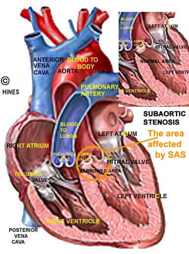
Aortic Stenosis/ Subaortic Stenosis AS/SAS
When renewed (oxygenated) blood leaves your pet’s left heart ventricle, it encounters a valve and a number of twists and bends as it exits. That path can be too narrow at many points or the valve (the aortic valve) can be poorly developed and unable to open fully enough to allow blood to pass freely. In some cases, an abnormal fibrous band, ring or ridge is present below the valve that interferes with blood flow and valve action. That forces your pet’s heart to work harder pushing blood through the narrowed passage. That added work eventually causes a heart to begin to fail. These problems are considerably more common in dogs than cats. In cats, they can be confused with, or perhaps even lead to hypertrophic cardiomyopathy.
SAS is more common in large breeds of dogs than small ones. Puppies prone to these problems can be identified as early as 4–8 weeks. Veterinarians don’t commonly associate AS/SAS, with runts or retarded growth – in most cases, the health issues that this problem causes take longer to develop. But in human infants, it’s associated with retarded growth and a few veterinary centers (like UPEI) mention seeing a link between the condition and slowed puppy growth.
You can read an article of mine on aortic/subaortic stenosis in dogs here. The end result in moderate to severe cases is congestive heart failure and the medical treatments for that in the young or the old do not differ. You can read about that here.
Current surgical techniques that veterinarians have available to deal with SAS are considerably less successful than those for patent ductus arteriosus. So, dogs and cats are generally managed with medications. However, a veterinarian with the Cardiology and Interventional Medicine department at Ohio State University has adapted a balloon valvuloplasty technique in an attempt to correct these problems by stretching the areas involved. This is done through a catheter placed in the pet’s neck or groin (A vet in Bristol, UK uses a similar technique, and I am told that the University of Florida has also attempted the procedure – perhaps also at Davis, CA). New techniques take time to perfect and there is still disagreement through 2016 as to the long-term benefits of the stretch procedure. That probably differs greatly on a case-by-case basis (perhaps longer-term success will require a fine wire mesh [stent] as seen in the video). Veterinarians with these highly advanced technical skills usually hang their hats in university “interventional radiology” departments).
In many cases of AS/SAS, owners see no initial signs or symptoms that the disease is present in their puppy; but the tests your veterinarian will run to detect it will be the same as those I mentioned for PDA.
There are also genetic tests to help locate dogs that carry the genes involved in SAS. I believe they are available through North Carolina State University Vet School and the Vet School in Davis, CA.
Pulmonary Artery Stenosis = Pulmonic Stenosis, PS
Your pet’s oxygen-poor blood can encounter obstructions on its way to the lungs as well. Its pulmonary artery and its tributaries (peripherals) deliver oxygen-poor blood returning from its body to the lungs for re-oxygenation. On its way there, it encounters the pulmonary valve. Some puppies and kittens are born with this channel too narrow at one point or another. In many cases, it is the valve itself (the pulmonary semilunar valve) that functions improperly (valvular dysplasia). In other rarer cases, the blood channels themselves are too narrow. (read here)
A heart murmur is usually present when the restriction is significant. And, depending on the degree, that blood flow to the lungs is hindered, and the heart can slowly fail due to the abnormally high local blood pressures (pulmonary hypertension) these restrictions generate. In mild cases, where blood flow restriction is minimal, no outward signs might be seen or the pet might just tire easily. In severe cases, the heart enlargement can cause abnormal heart rhythms (arrhythmias) that lead to sudden death. In others, blood fluid, leaks from swollen veins and floats free in the abdomen, distending it (ascites) or pools under the skin of the legs (pitting edema).
Dogs with flat faces (like boxers, Bostons, Bulldogs, etc.) are often associated (over-represented) with this problem. But many other breeds are occasionally affected. Puppies and kittens that have severe forms of this defect often fall behind in their growth. It is not that unusual for more than one heart defect to be discovered during your pet’s diagnostic workup. (read here)
What Can Be Done?
The same “minimally invasive” stretching technique I mentioned, balloon valvuloplasty, has been used to treat pulmonary valve stenosis in puppies and kittens. The results are mixed. They seem to give the best results when the leaves (valve flaps) of the pulmonary valve are incompletely separated and the stretching apparatus is capable of forcefully separating (tearing) them apart. Not all cases are suitable for this technique.
Are There Medications That Might Help?
Some veterinary schools suggest that pets with this problem can be helped by drugs like atenolol that lower blood pressure. But humans with pulmonic stenosis are most commonly given sildenafil (Viagra®), tadalafil (Cialis®) or ambrisentan (Letairis®), newer medications that lower pulmonary blood pressure in a different way. Experimentally, ambrisentan is quite effective in lowering blood pressure in dogs. But I do not know of it having been suggested for this problem. I did have one case in NY where sildenafil was dispensed. The ProBNP test and echocardiography might help your veterinarian decide if and when those medications might be beneficial.
Persistent Right Aortic Arch One Of Many “Vascular Ring” Birth Defects or Anomalies

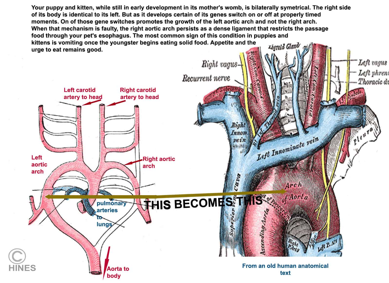
When your dog or cat was a small, primitive embryo, it had six paired aortic arches. Some were destined to fade away when their tasks were complete; others to later become portions of the youngster’s major blood vessels. One of the fourth pair was was destined to become the portion of your pet’s aorta that curves over the forward portion of its heart.Occasionally, that process does not occur normally. In some cases, it forms in a mirror image to what it should be, in others, bands of fibrous, ligament-like tissue remain that form a tight ring around the youngster’s windpipe and esophagus. As the dog or cat continues to develop, these bands do not allow food to pass through the ring of tissue. The defect is particularly common in German shepherd dogs and German pinschers (read here) ; but it can develop in any breed. It is a considerably rarer birth defect in cats.
These youngsters usually appear normal until they began to consume solid food – that is because milk can pass through a much narrower channel. Then, as they switch to a solid diet, they eat – only to throw up the undigested food shortly thereafter. Needless to say, their growth falls behind. (Not all German shepherds that develop these signs have a persistent right aortic arch. Some of those that develop it later in life have megaesophagus or other genetically related causes.
A barium swallow is the most effective way to diagnose an esophagus blockage or motility problem. Barium shows up well on x-rays, and it accumulates just ahead of the stricture (or blockage) – as in this image: 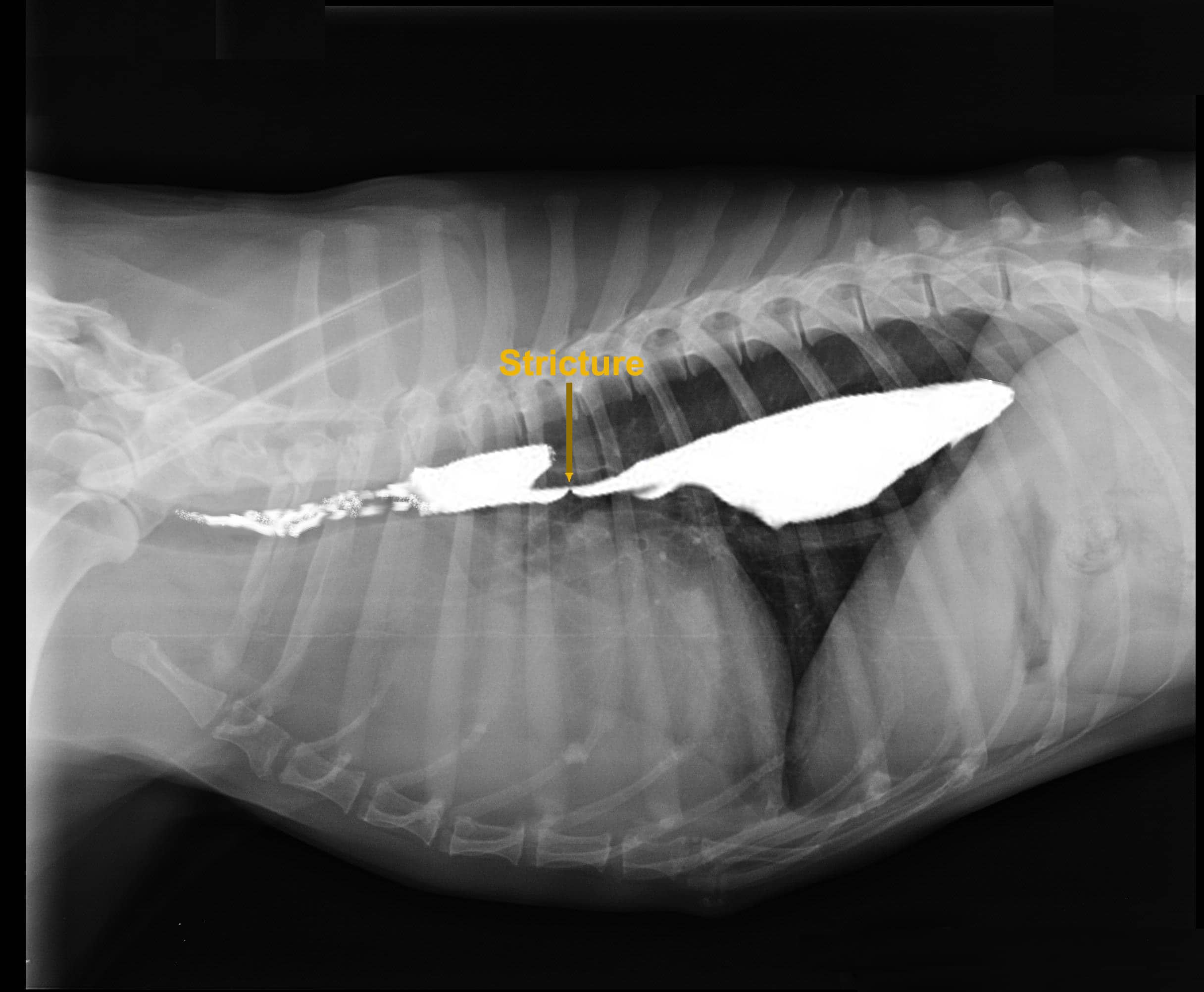 A barium slurry, being a liquid with the consistency of cream, passes. But sold food has difficulty doing so. Large institutions give the barium while viewing its real-time progress with a fluoroscope, smaller facilities take a series of x-rays like the one pictured (if the facility has ways to visualize the movement of the barium in real time, the lack or presence of normal esophageal muscle contractions is also apparent. That tells your vet more about your treatment success likelihoods).
Many of these cases can be helped surgically by cutting the fibrous band (the ring or stricture) and allowing the esophagus to expand to its normal diameter. However, in many pups or kittens that have had this problem for an extended period, the esophagus is so greatly stretched into a sac (= megaesophagus) ahead of the stricture that it does not regain its normal function. In others, some function returns, but the pet does best eating with its head and chest elevated (e.g. food dish placed on a chair, food moist, food blender, etc.). When full recovery occurs, it can take time. Some cases of megaesophagus treated later in life have aortic arch problems as their underlying cause. (read here) Other cases of megaesophagus that hinder eating are due to birth defects in the nerves that regulate esophagus motility. (read here)
There are many more heart defects that your puppy or kitten might be born with that can affect its growth rate and lifespan. Fortunately, they are quite rare. Some can be corrected, many cannot. They include defects (holes) in the heart chamber walls (atrial and ventricular septal defects) tetralogy of Fallot, imperfectly formed mitral or tricuspid valve (tricuspid valve dysplasia in kittens), endocardial fibroelastosis of cats, cor triatriatum sinister in cats, etc. When serious, all of them cause easy fatigue, shortness of breath, and often, stunted growth. The early signs are due to the youngster not receiving enough oxygen because blood does not move through its body normally. The later signs are the signs of an enlarging heart that is in failure. Many, but not all, produce heart murmurs. Some produce abnormal heart rhythms (arrhythmias) and an abnormally rapid heart rate. When not surgically correctable, heart failure can usually be delayed with a cocktail of medications – drugs to allow the heart to work less (ace inhibitors, beta blockers PDE5 inhibitors) those that increase the strength of its contractions (positive inotropes). Diuretics, like furosemide and spironolactone, help to clear excess pooled fluid from the body.
Portosystemic Shunts
Blood circulation errors can occur farther from the heart. A common location is in or near the puppy or kitten’s liver. How blood moves through the liver is critical for processing the nutrients that arrive there from the pet’s intestines as well as in detoxifying and excreting the waste products of metabolism. I wrote an article specifically on portosystemic shunts. You can read it by clicking on the puppy and kitten above.
Portosystemic shunts occur more often in pure breed than mixed breed dogs – but both are occasionally born with them. The problem also occasionally affects kittens. Those kittens tend to be small for their age. Although portosystemic shunt problems are an inherited genetic problem, how it is inherited and which youngster has it is quite complex.
Hepatic Microvascular Dysplasia & Portal Vein Hypoplasia = PVH-MVD aka hepatoportal microvascular dysplasia” (HMD), Portal Atresia

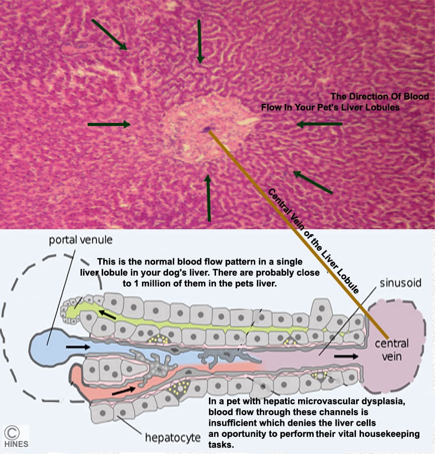
I devoted a small portion at the end of an article on portosystemic shunts (read here) to microvascular dysplasia of the liver. A problem most commonly affecting toy dog breeds. I now see that I should have discussed microvascular dysplasia at greater length because many more small dog owners write to me about their little dog facing microvascular dysplasia issues than do about portosystemic shunts, so I devoted some time to an article specifically on MVD. You can read it here
Other Circulatory Problems
It is not just blood that circulates throughout your pet. Lymph does too, and the lymphatic vessels are also subject to malformed, misdirected or missing lymph channels. These conditions are quite rare. But they have been reported in puppies and kittens.
Primary Lung Issues

There are kittens that are occasionally born with lungs that are not efficient in obtaining the oxygen they need. It is quite difficult – if not impossible – to tell those infants from those that are gasping or breathing with labor due to the other circulatory problems I discussed earlier. We do know that some of these infants have problems breathing because they lack complex protective compounds that normally line their lung spaces. Those compounds are called pulmonary surfactants.
Lung surfactants prevent the infant’s lung from collapsing too fully when it exhales. They also make it easier for oxygen in the lungs to enter the blood stream. They reduce surface tension.
Very little has been published on the dynamics of this problem in dogs and cats. But in human infants, we know that many things can cause it. In some cases, the genes that allow the body to produce them are defective. In others, it was a premature birth that caused the problem. In still others, inhalation of meconium while still in the womb seemed to be the cause. (read here & here)
When the problem is a genetic defect, there is little that can be done. But when it is due to a difficult birth, perhaps ACE inhibitors like enalapril might be of some help in getting it through its initial crisis. (read here & here)
Thyroid Gland Issues Hypothyroid Dwarfism
A thyroid gland that produces normal amounts of thyroid hormones is essential for a puppy or kitten’s normal growth. When a youngster’s thyroid gland cannot do that, one result will be dwarfism.
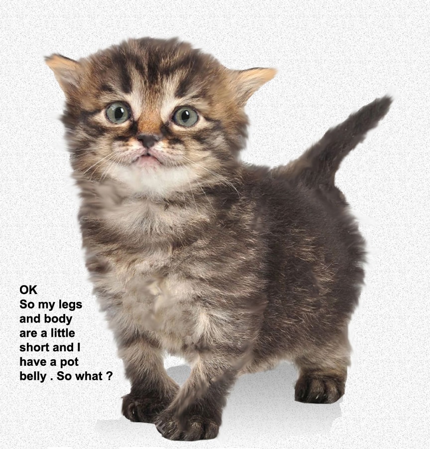
Youngsters that share that problem have a particular body shape. Their legs are short for the size of their body and their head and neck larger and broad. Their hair coats tend to be fine and soft, and they are often slow to develop their permanent teeth. Viewed from the front, their elbows often protrude from the midline (abducted). Constipation problems are common (which adds to their potbelly) and they tend to be abnormally calm, reserved, less responsive and attentive than their normal littermates. As these youngsters mature, veterinarians see a variety of skeletal abnormalities in them because proper bone development and bone maturation sequence also depend on adequate thyroid hormone.
Your puppy or kitten’s thyroid gland is under the control of its pituitary gland. So, the cause of this problem can be within the pet’s thyroid gland itself or within its pituitary – the master gland at the base of its brain. Abnormalities in the thyroid gland itself are varied. Sometimes the gland lacks the ability to manipulate the iodine needed to form the thyroid hormone (organfication = can’t produce TPO). In other cases, the thyroid gland did not form completely (agenesis/dysgenesis) while the infant was still a fetus (remember, at that time its mother’s thyroid hormones were carrying its development process along). In some, the cells that produce thyroid hormone lack the ability to respond to pituitary TSH hormone commands to produce thyroid hormones (TSH unresponsiveness). (read here & here) These were all examples of Primary Hypothyroidism – that is, the problem was within the youngster’s thyroid gland itself. Here is the second form:
Secondary Hypothyroidism ….When The Pet’s Pituitary Gland Is Really To Blame
As I mentioned, the youngster’s thyroid gland gets its orders from the pituitary gland in the form of thyroid stimulating hormone (TSH aka thyrotropin). TSH instructs the thyroid to produce T3 and T4 hormones. If those signals do not come, no T3 or T4 will be produced. One of the many results of that will be a smaller than normal youngster. When the puppy or kitten has pituitary gland problems, it is not uncommon for other endocrine glands in their bodies to also be under active. You see, the pituitary gland does not only produce the TSH that drives the thyroid gland, it also produces the hormone ACTH that stimulates the pet’s adrenal glands to produce cortisone, FSH and LH (gonadotropins) that are essential to proper ovary and testicle maturation as well as growth hormone (GH) that drives normal growth. When the pituitary fails to produce multiple hormones, the problem is called combined pituitary hormone deficiency (CPHD) or panhypopituitarism. In those cases, stunted growth can be due to a combination of low thyroid hormone and growth hormone as well as other health issues related to CPHD.
How Can My Vet Confirm That This Is The Problem?
Besides confirming that the typical symptoms of hypothyroidism are present in your pet, your veterinarian will measure the amount of T4 hormone that the kitten or puppy’s thyroid gland is producing. The blood test is not foolproof in the very young – it can be low for non-thyroid reasons. If TSH is also low, suspicion falls on the pet’s pituitary gland as the root of the problem. However, the best test to confirm that is to give synthetic TSH to see if the pet now produces thyroid hormone normally. (read here) These tests are best sent to a university or national veterinary testing laboratory. That facility must be informed that the sample is from a very young pet because normal T-4 values in adult dogs and cats are quite different (usually lower) than normal values in the very young.
If your veterinarian wants more help in deciding if the underlying problem is in the youngster’s thyroid gland or in its pituitary gland, he/she might decide to check the pet’s IGF-1 (insulin-like growth factor-1) level. But because that level in pets is said to fluctuate greatly during the day, the results of a single test can be of minimal value. In humans, low values suggest pituitary issues as well; but in us the test results are just as hard to interpret. Persistent low values point to the pituitary gland as the underlying problem.
What Can Be Done For A Hypothyroid Youngster?
Congenital hypothyroidism, like adult-onset hypothyroidism is quite treatable. Drugstore levothyroxine– which is the same as the T4 your pet lacks – can be given as an oral supplement. But success depends on the age at which treatment is started because many of the effects of a lack of thyroid hormone in the youngster’s growth period cannot be reversed. Your vet will want to fine tune the dosage based on the pet’s periodic blood T4 levels. Re-read some of the links I gave you before for more details.
Are There Other Genetic Problems That Might Be Mistaken for Thyroid or Pituitary Dwarfism?
Yes.
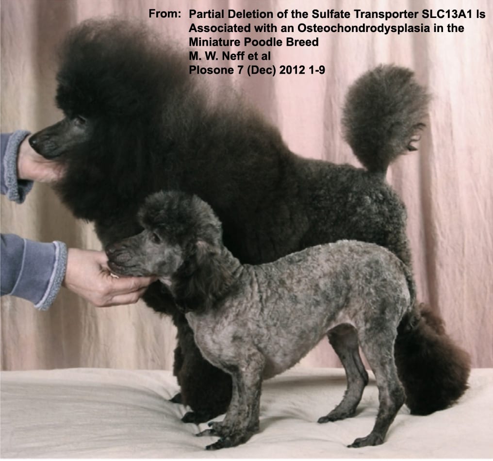
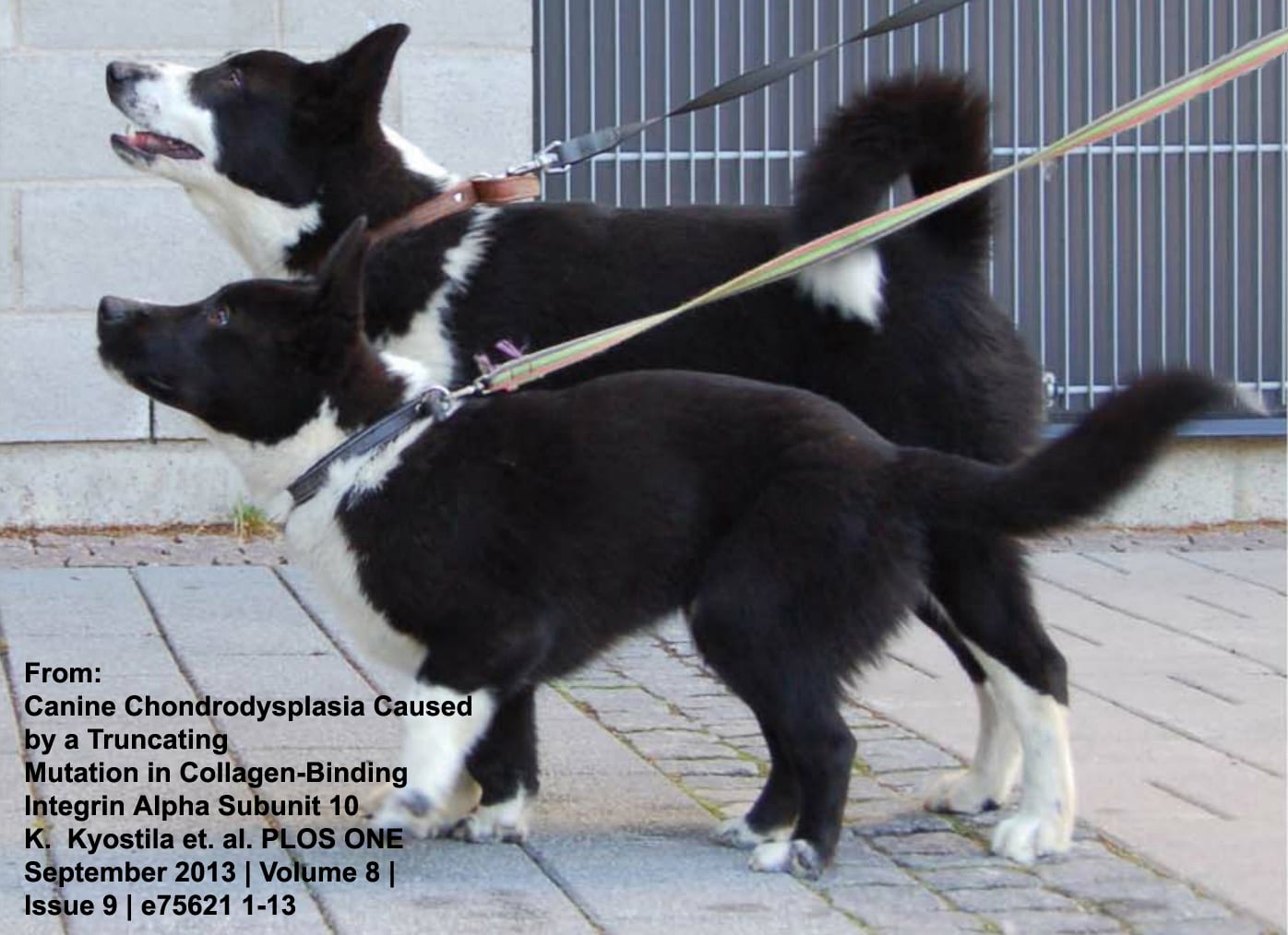
I am sure that many exist and that the causes of many have not yet been discovered. The smaller of the two poodles above was born missing a crucial gene that allows it to absorb sulfur (sulfate) from its food. Consequently, it could not produce important sulfur-containing amino acids. The smaller of the two Karelian Bear Dogs in the second photo has a genetic skeletal disorder called chondrodysplasia.
Normal bone formation is a complex process that can be interrupted by a genetic defect at many points.
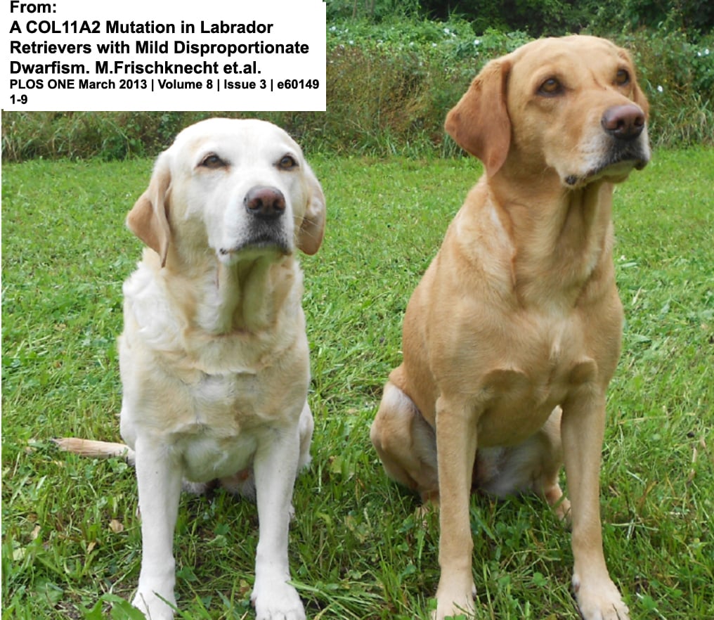
Some growth defects, like the one above, affecting the slightly smaller of the two yellow Labradors, are quite subtle. That defect produces pups that are healthy, but with legs disproportionately shorter than their parents. As is the case in many genetically based diseases, the single rare gene mutation that causes the problem was unknowingly spread when a normal-appearing carrier dog was used for breeding.
So as Hines harps about in this and other articles, beware the “rare”, the “unique” and the “striking”, the “exotic” and “trendy”, and the “new emerging breed” when selecting your kitten or puppy. That pet or one from “a special line of show champions” or anything else that hints of consanguinity and inbreeding can also be unique or special in ways you had not anticipated. In cats, even things like increased susceptibility to FIP can be a result. (read here)
Growth Hormone Deficiency
I mentioned that when the puppy or kittens small size is related to defects in its pituitary gland, it could be the combined effect of a lack of thyroid stimulating hormone or a lack of growth hormone or a lack of both that keeps it small. Looking through published reports, I believe that when a puppy or kitten’s pituitary gland is not cranking out its normal hormones, it is most common for it to produce insufficient amounts of several different hormones, not just one kind.
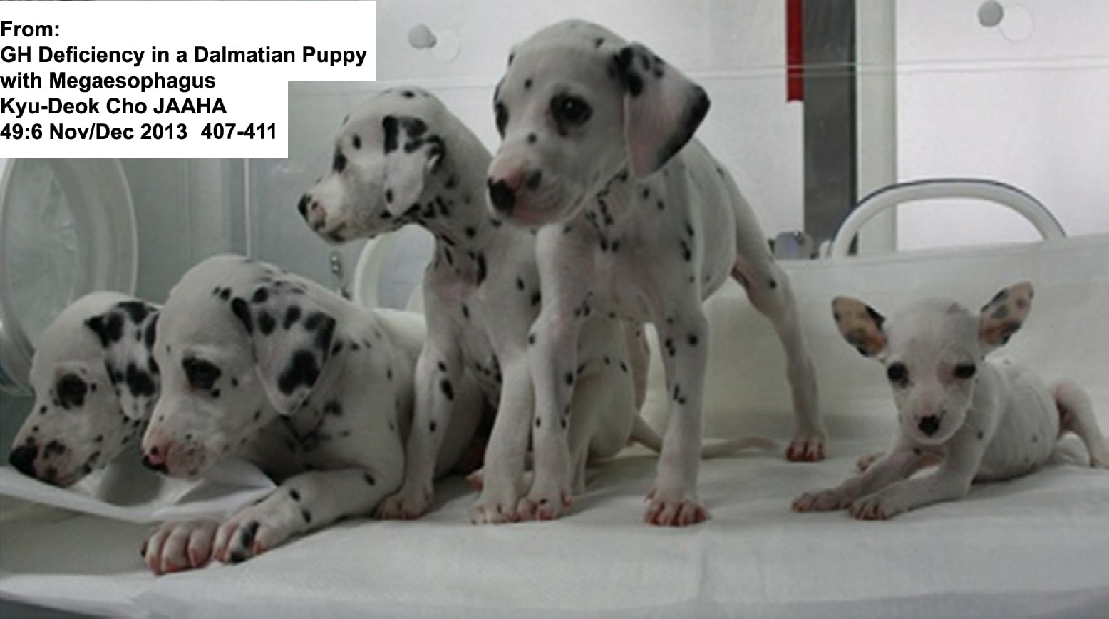
But that is not always the case – as in the photo above. You see, just as the thyroid gland relies on a signal hormone (TSH) from the pituitary gland to begin producing thyroid hormones, the pituitary gland relies on another signal hormone (GHRH) produced higher up in the brain to stimulate the youngster’s pituitary gland to release growth hormone (GH). Now if the receptors for that signal hormone (GNRH) in the pituitary gland of the pet are genetically defective, no growth hormone (GH) will be produced by its pituitary gland. That sort of situation occasionally occurs, and it leads to only a growth hormone deficiency. It is called hyposomatotropism (somatotropin is an alternate name for growth hormone or GH).
I do not know if that problem has been confirmed without a doubt in puppies or kittens. But it has been in mice and humans. There is no reason to believe that it does not occasionally occur in dogs and cats as well. The one report that I know of concerning a possible case of hyposomatotropism in a dalmatian puppy is the one in the photograph. I am uncertain if sufficient tests were performed to completely rule out that the pup did not have a thyroid stimulating hormone deficiency (TSH) as well.
Genetic Defects In Your Puppy or Kitten’s Basic Metabolism
Your puppy or kittens intricate metabolic pathways are under the strict control of its genes. Some of those genes regulate the absorption of nutrients, others determine how nutrients are processed or how waste products are detoxified and eliminated; and still others, how and where metabolic products are stored. Critical to performing many of those duties correctly, are the lysosomes within your pet’s individual cells. Two common genetic defects affect the lysosomes of dogs and cats, and humans as well:
Lysosomal Storage Disease aka Lipid Storage Disease
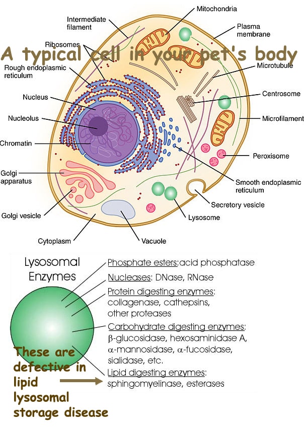
Most cells in your pet’s body contain lysosomes. They are little packets (vesicles) of enzymes that digest the complex molecules that make up the pet’s body into their basic units for reassembly into other compounds or prepare them for elimination from the body – somewhat like the stomach – but on a much smaller scale (which is why they are called organelles).
Every enzyme in the lysosome is produced under the direction of a pair of genes. If one of those gene pairs is defective, the lysosomes cannot digest the particular substance under that enzyme’s control and that substance can build up within the cell – eventually killing it.
One of those enzymes (beta-galactosidase) is required to process fatty substances (lipids) found within cells. Those fatty substances are particularly common in the nerve cells that make up your pet’s nervous system. So pets inheriting this disorder may appear normal at birth and for several months thereafter, but gradually they loose coordination and other nerve function throughout their bodies. Some remain abnormally small in size as well (proportional dwarfism).
This problem has been seen in English springers, Portuguese water dogs, beagles and huskies as well as kittens – both purebred and mixed bred dogs can, on occasion, suffer from this defect.
Mucopolysaccharidosis ….Another Form of Lysosomal Storage Disease
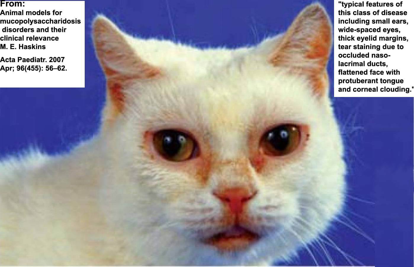
You can read the article from which this photo comes: (read here)
In the lipid storage form of lysosomal storage disease, I mentioned that dogs and cats can be born with decreased or missing lysosome enzymes that are necessary for them to process fatty substances (lipids).
But dogs and cats can also be born missing other lysosome enzymes that would normally allow them to process complex sugars too (long chain carbohydrates). One of the principal long chain carbohydrates that causes problems when it builds up due to this genetic defect is the same one vets sell in an attempt to control arthritis, glycosaminoglycan (GAG), a constituent of chondroitin. Over time, this compound builds up where it does not belong and damages various organs in the pet’s body.
Pets born with this problem are often smaller than normal at birth. It is not unusual for them to have a somewhat distorted face and/or eye defects (corneal opacities). Their gait (how they walk) is often abnormal. Their limbs can be distorted, and they tend to fall behind their littermates in their ability to move about. Many other abnormalities, located throughout the body, are also common when pets harbor this genetic abnormality. Because chondroitin is a major contributor to the strength of cartilage and the disease interrupts where the material is normally laid down, puppies and kittens with this disease tend to have floppy ears and lax (relaxed) joints and, perhaps, a predisposition to hernias. That abnormal buildup of chondroitin in the pet’s corneas is also one of the reasons for their cloudy eyes. You can read two of the four key articles that describe mucopolysaccharidosis in puppies and kittens here: (read here & here) Ask me for Dombrowski2004 & Haskins1979 if you want the other two.
Can My Vet Confirm This Problem?
Yes
However, the diagnosis is not a thing that can be done during an ordinary office call. It requires a veterinary pathologist’s careful examination of stained blood film slides from the pet. If mucopolysaccharidosis is the underlying problem, some neutrophil and lymphocyte cells in the blood film generally contain large numbers of small purplish granules (azurophilic granules). The granules consist of the excess glycosaminoglycans circulating in the youngster’s body.
Those glycans also spill into the pet’s urine in excessive amounts. A test exits to see if their level in the urine is excessively high.
Since it is so rare to run the test on pets and because there are multiple variations of the disease, it is highly desirable to compare the results to a normal littermate.
Is There Anything My Vet Can Do About It?
Currently, all that can be done is to make the pet as comfortable as possible and treat its various disabilities symptomatically as they occur. Experimentally, gene therapy, bone marrow transplants and enzyme replacement seem to have helped some. (read here, here & here)
Clefts
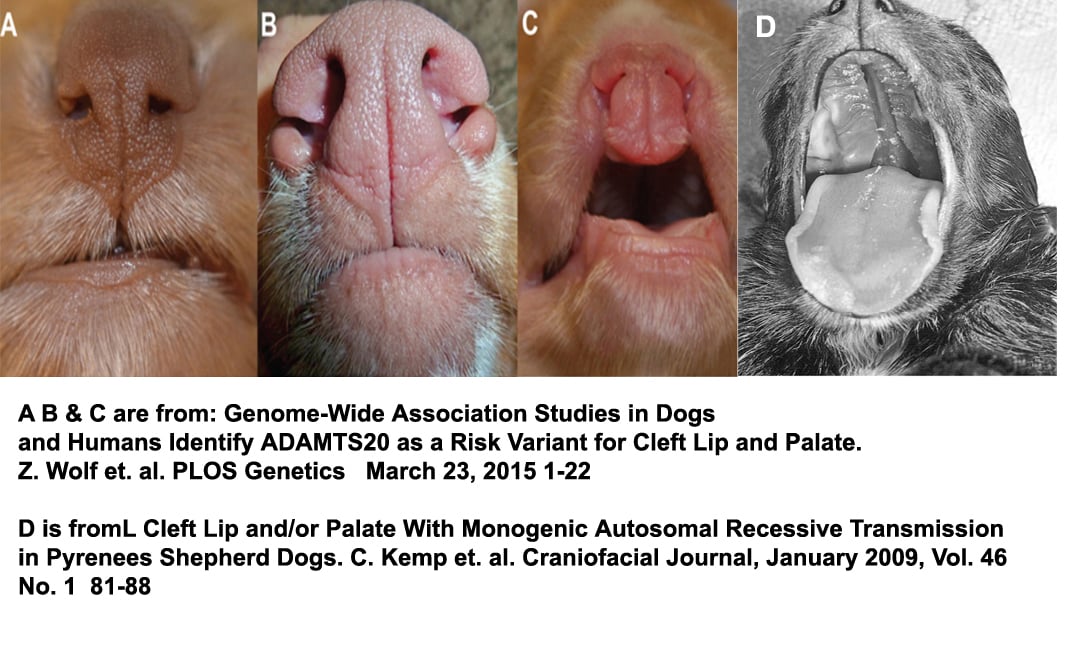
Some gene defects prevent cells from carrying on vital functions. But other gene defects affect how cells self-organize in the embryo while in the womb. Many of those are signaling genes that are meant to reshuffle the body’s shape as it makes the tremendous changes required for independent living (like the ones I mentioned as the cause of aortic arch problems). When that process goes wrong, kittens and puppies can also be left with openings (clefts) that were essential during fetal development but life-threatening when they do not close at the proper time. It is not that unusual for youngsters with clefts to have other developmental defects as well. Veterinarians do not know what percentage of cleft lips and cleft palates (orofacial clefts) are due to defective genes and what percentage are due to environmental events that occurred during pregnancy. (read here & here) One or both can be in play in any particular case. However, there are breeds that have a known genetic susceptibility to clefts (e.g. Nova Scotia Duck Tolling Retriever and Pyrenees Shepherd Dogs) where specific defective genes are almost certainly the cause. When veterinarians don’t know if the problem was genetic or womb-environment caused, they just call it a congenital disease.
Puppies and kittens with facial clefts appear to be more susceptible to middle ear and respiratory infections. That can lead to deafness, among other things. In cases that involve the floppy soft pallet, that organ may simply be smaller than it should be and not able to entirely block food from entering the windpipe and lungs rather than the esophagus while the pet is eating. Chronic nasal drainages (rhinitis/sinusitis) are also common.
Often, the first sign of the problem that pet owners notice is that their kitten or puppy has milk or snot coming out of its nose.
Facial Deformities:

The upper and lower jaws of cats and dogs are genetically programmed to grow at the same rate. Desired gene mutations are assumed to have caused this not to happen in the flat faced breeds such as boxers, bulldogs and Shih Tzus. The words prognathism or its reverse, brachygnathism, are used to describe that appearance. More commonly, it is called an under bite or an undershot jaw or the reverse, an overbite or overshot jaw when it is the lower jaw that is shortened (as seen occasionally in dachshunds and beagles). Either an overshot or an undershot jaw can make eating difficult when the misalignment is severe – but more importantly, misaligned upper and lower canine teeth can cause painful gum sores.
The problem is considerably more common in puppies than kittens. This deformity does not, in itself, cause stunted growth. However, it is occasionally part of multiple genetic problems that do. (read here, here, here & here)
Skilled veterinary surgeons can correct the condition in a youngster once it has grown to its full adult size. But lifestyle changes and dental work are usually sufficient to give these pets a comfortable life.
Hydrocephalus
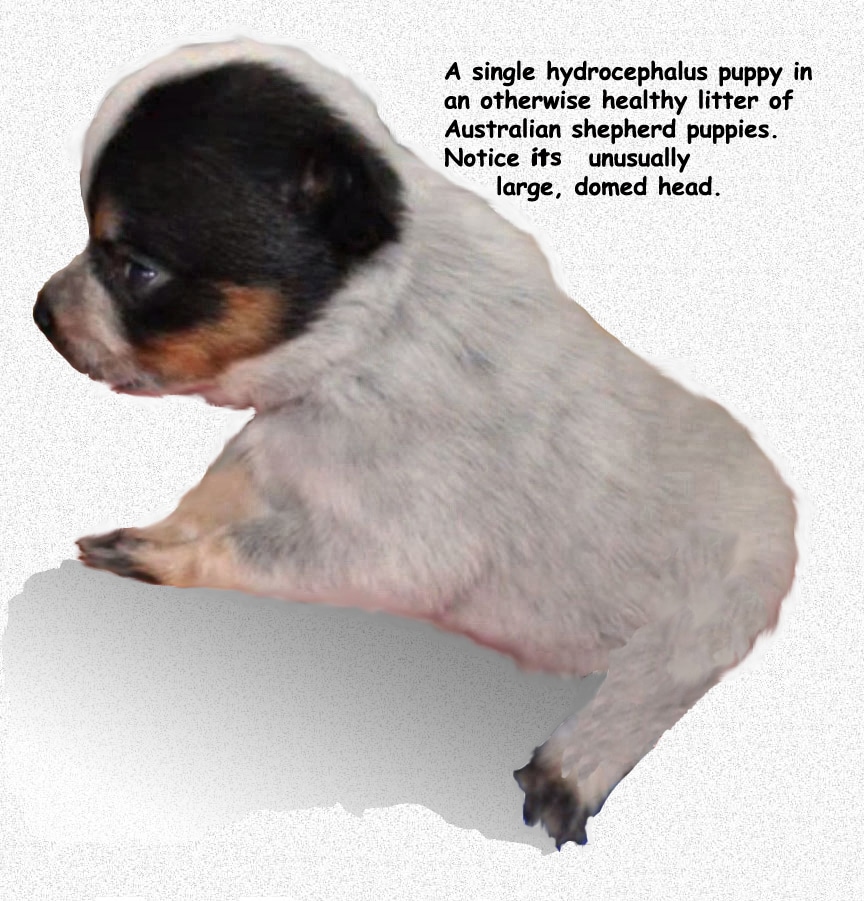
Your kitten or puppy has a series of fluid-filled chambers within its brain that persist throughout its life. Just like the lower chambers of the heart, they are called ventricles. There are four in the brain. They are filled with cerebral spinal fluid (CSF). All chambers interconnect. A channel leads down and surrounds the entire length of the pet’s brain and spinal cord as well. The fluid in the chambers is normally under low pressure (much lower pressure than the pet’s blood pressure – 8-9 mm Hg vs 140 mm Hg systolic blood pressure in the dog).
Cerebrospinal fluid is in constant slow circulation in the system as it bathes and protects the brain, delivering and removing various elements. That flow can be interrupted by blockages at various points in the system. When that occurs, CSF pressure increases, destroying brain tissue and causing the fluid-filled chambers to enlarge. As the brain enlarges, the top of the head that encloses it expands as well. The end result in puppies and kittens is hydrocephalus.
Many of these infants are born smaller than their littermates – probably because of other genetic defects that often accompany the problem. In others, the infant is of normal size with only an enlarged, domed or apple-like heads at birth and does not begin to lose its normal behaviors until CSF pressure rises higher as it approaches weaning age. That is because as brain tissue is slowly lost, mental capacity is lost as well (ie cognitive dysfunction). That can progress to blindness, depression, head-pressing, random circling, blindness, seizures and death.
In cats, intrauterine infection with the panleukopenia virus is one suspected cause, in others, certain medications given to the queen (e.g. griseofulvin) can be to blame. Since it is seen most commonly in purebred cats (particularly Siamese) and the toy breeds of dogs, veterinarians believe that there is a genetic basis as well. But how genetics and environment interplay to produce hydrocephalus remains unknown. In mild cases, changes are not dramatic. A common complaint in those situations is owner frustration at not being able to house train the pet.
How will my Vet Be Sure That Hydrocephalus Is The Problem?
The majority of hydrocephalus cases are diagnosed based on the head conformation of the youngster in relation to its littermates as well as the mental problems the youngster is experiencing. If owners want a more positive diagnosis, the sizes of the brain ventricles in relation to their brain size need to be measured. CAT scan and MIR examinations are the best way to do that, although an ultrasound examination can be helpful when the bones of the skull have not yet fused. When the fluid-filled ventricular spaces are only slightly or moderately enlarged, those examinations can be hard to interpret – particularly if only a single pup or kitten in the litter is available for contrast with its normal littermates.
In many cases, the central soft spot (fontanelle) at the top of the infant’s skull will be larger than normal. Breeds that normally have an apple or dome-shaped heads can make the diagnosis in those breed very challenging.
Are There Things My Veterinarian Can Do For Those Kittens and Puppies?
Whatever brain damage that has occurred is not reversible. But further brain damage can sometimes be limited by placing a tube (ventriculoperitoneal shunt) from the fluid-filled chambers to the pet’s abdominal cavity to serve as an alternative channel for the cerebrospinal fluid to leave the brain. The brain is remarkable in its ability to adapt and re-rout thought processes. So, the pet’s general demeanor often improves, and its limitations diminish once the pressure problem is solved. It is a difficult operation and side effects and failures are not uncommon. It is a procedure best left to an advanced veterinary teaching facilities.
Medications can sometimes also slow the progress of the disease. Certain diuretics (e.g. Lasix®/furosemide, acetazolamide/methazolamide) decrease the production of cerebrospinal fluid and have been successful in decreasing cerebral spinal fluid pressure and reducing any brain swelling it has produced. Omeprazole (Prilosec®) combined with corticosteroids (prednisone/dexamethasone) have been successful in reducing inflammation that may be blocking normal CSF drainage. Seizures can often be reduced or eliminated with the same medications veterinarians use to control epilepsy. The earlier surgery is attempted or medications given, the more likely they are to be beneficial.
Digestive Tract Issues – Swallowing difficulties Megaesophagus
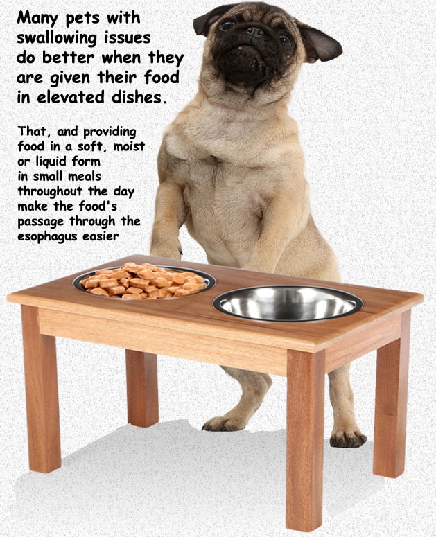
Some puppies, and an occasional kitten, will develop swallowing problems. They generally become noticeable shortly after the youngster begins eating solid food. Besides limiting their growth, these youngsters are at much higher risk for pneumonia because the brought up (regurgitated) food can enter their lungs. The symptoms are the same as the symptoms of a retained aortic arch that I discussed earlier. But in this case, the juvenile’s esophagus is not constricted by a fibrous ring, instead, the nerves that normally stimulate the esophageal muscles to constrict in rhythmic waves are dysfunctional. The problem can also occur as a secondary megaesophagus when a fibrous esophageal scar of any sort makes the passage of solid food difficult. It can also occur as secondary megaesophagus when the sphincter (muscular valve) at the base of the esophagus (nearest to the stomach) fails to open normally to allow the passage of food (achalasia). Hypothyroidism has also been identified as an underlying cause in some cases.
These pets tend to have good appetites. But a few minutes to a few hours after eating, the undigested food is brought back up (often with an attempt to re-eat it again). The youngsters become lean or quite thin due to malnutrition unless their food is liquefied or given as a slurry.
When veterinarians suspect this problem, the pet is generally given an oral dose of barium sulfate suspension (a barium swallow) that allows the veterinarian to visually follow the passage of the barium material through the esophagus on its way to the stomach. When a stricture due to a persistent aortic arch or, perhaps, having fed scalding milk replacement are ruled out; and much rarer diseases such as myasthenia gravis, or hypoadrenocorticism are also been ruled out, primary megaesophagus is the most likely cause. There are some cases of primary megaesophagus in which youngsters improve on their own as time passes. (read here) I have a better, more inclusive article, that I cannot include here; but if you ask me for Mace2012.pdf.
Incomplete Formation of The Digestive Tract and posterior spine
 So why the hedgehog? It’s to remind us that the sonic hedgehog signaling pathway is thought to play an important role in many of these defects.
The most common location for a portion of a puppy or kitten’s digestive tract to be lacking is the youngster’s anus and the portion of the rectum just before it. When only the anus is lacking or abnormally formed, it is often called atresia ani (aka imperforate anus. Because there is no limit to how many ways these types of missing portions can occur – so many names describe what is basically the same phenomenon).
In some cases, there is complete lack of exit from the pet’s large intestine, in others a thin membrane blocks passage and in still others the exit is smaller than it should be (stenotic). Even the anal sacks (the smelly secretion glands) on either side of the pet’s normal anus can be missing or imperfectly formed. In those severe forms, it is also common for the muscles that normally keep the anus closed between bowel movements to be missing as well. In females, the rectum sometimes communicates with the vagina (vaginal fistula) – with feces exiting that rout. Many can be corrected with surgery – some through the use of devices that expand constrictions (dilator bougies). But a portion of the animals do not regain fecal control.
It is common for breeders and owners not to recognize the problem until the infant becomes bloated and begins to strain due to fecal impaction. Although these problems are relatively rare, poodles, Boston Terriers and English bulldogs seem to develop them in a higher numbers than other dog breeds (read here); Manx and other bobtail cat breeds as well. When the problem is present, their growth is often stunted – perhaps due to multiple congenital malformations and omissions. (read here, here & here)
Veterinarians really do not know how many of these sort of birth defects that they see are due to defective genes and how many are due to environmental factors within the mother’s womb. When defective genes are implicated, both hedgehog and another group, the T-box gene family are prime suspects. (read here).
Similar problems also occur in human babies. Different genes regulate our development at different stages of our fetal life. They are known to interplay with the mother’s womb environment in occasionally causing these unfortunate problems.
Neurological Issues
It can be quite difficult to determine if newborn puppies or kittens have defects in their nervous systems. That is because youngsters are not capable of coordinated movements – other than those associated with nursing – until they are 7–10 days old. It is generally not until their third week that their legs can support their body weight. Even then, they are quite uncoordinated until they are about 18–21 days old. Kittens develop these skills a bit sooner than puppies, but still, it is not until about 14–28 days that the permanent incoordination (ataxia/cerebellar hypoplasia) related to a panleukopenia virus infection they obtained while in the womb becomes apparent in kittens. When similar signs occur in puppies; the majority of them are due to defective genetics. (read here, here, here & here)
Coordination Problems – Ataxia and Tremors
Any portion of your pet’s nervous system is subject to defects in its development due to deficiencies in its womb environment, nutritional deficiencies in the mother or genetic errors. But coordination problems are the ones that veterinarians recognize most.
Your puppy or kittens ability to move smoothly relies on a portion of its brain called the cerebellum. I mentioned that a panleukopenia (the parvovirus of cats) infection in a pregnant mother cat and its unborn offspring (a prenatal infection) is the most common cause for this lack of coordination in kittens. That is because parvovirus prefer to infect cells that are dividing rapidly, and the cerebellum, at that stage of development meets the virus’ needs well. Inborn genetic defects have the ability to cause similar signs, but those cases are considerably less common in cats. (read here) How virus-induced cerebellar hypoplasia influences a kitten’s growth rate is highly variable – even in kittens from the same litter. Mild cases may just walk mechanically – like a windup toy – and occasional head tremors (intention tremors). But more severely affected kittens can only walk and stand with great difficulty. In unsupervised and unassisted situations, those kittens’ growth rate will be retarded. In other cases the neurological symptoms are considerably more severe. (read here)
Old English Sheepdog, Gordon setter, Scottish terrier, Staffordshire terrier, spinone iltaliano and Finnish hounds occasionally suffer from a different genetic defect that produces symptoms quite similar to cerebellar hypoplasia in kittens. That hereditary disease can begin in young puppies, but it can also occur later in life. How progressive the disease will be and how individual puppies will be affected is quite variable.
Urinary Tract And Kidney Issues
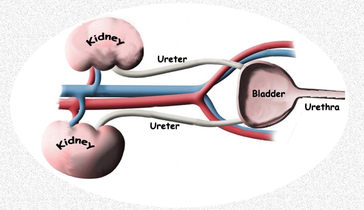
Genetic defects can also influence the development of your pet’s kidneys and urinary tract. In rodents and humans, defects in the same T-box gene family that I mentioned regarding missing or defective spinal components (read here) are often involved. (read here) Those genes are often lethal when two defective members of a pair (alleles) are inherited. So, puppies and kittens in those situations would most likely die before birth.
Defects can affect any portion of a pet’s urinary tract. Sometimes, kittens and puppies are born with incomplete or malformed tubing (ureters & urethra). Other genetic mutations, inherited from their parents lead to early kidney failure (read here) Another genetic defect causes early blockages with urinary tract (with stones=renal calculi). (read here & here)
It is not unusual for these star-crossed youngsters to have been born with multiple health issues that relate to various systems in their body besides their kidneys and urinary tract. That is because signaling genes often join with others to perform different tasks at different locations. If you have read some of my reference links, you can see that is often the case. It is also why the long-term outlook for a youngster with only one recognized defect so far may be guarded and why it might face other challenges farther down life’s path.
Polycystic Kidney Disease (PKD)
One of the most common and most serious urinary tract defects is polycystic kidney disease (PKD). Although the very early beginnings of the kidney cysts that will bring on this disease are present at birth, it is rare for them to cause obvious signs before the youngster is 6 months old and much more likely to begin considerably later in life than that. That is because it takes time for these fluid-filled cysts to enlarge sufficiently to destroy the normal kidney tissue around them.
Signs of the disease (uremia) (read here) are not noticeable until about 2/3 of your pet’s normal functional kidney tissue has been destroyed. Persians, Himalayan and British shorthair cats are the primary breeds affected. Polycystic kidney disease is less frequently seen in dogs. When it does occur in dogs, it is generally, but not always in a purebred. (read here)
There is no cure for polycystic kidney disease. But these pets can be managed, and time gained with the same techniques used in other forms of kidney failure. (read here)
PKD is said to be the most common genetically inherited disease of cats. It is an unusual one in that most genetic defects I discussed up to now require both of the two members of any specific gene pair to be defective in order for the disease to occur (= a recessive trait). Those with only one defective gene for a recessive trait are carriers of the problem; but it is only the next generation that is at risk if both parents carry one defective copy. Polycystic kidney disease is different in that even a single defective gene can trigger the disease. That means that the defective trait is dominant.
Kittens that inherited two defective genes in the pair (alleles) do not survive to term. The disease in cats is sometimes referred to as autosomal-dominant polycystic kidney disease or ADPKD.
Weak Immune System Issues And Infections
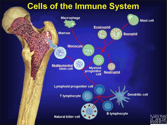
Your puppy or kitten’s adaptive immune system did not come strongly into play until several weeks after its birth. Until that time – and during the period it was sheltered in the womb – it relied on the adaptive immune system of its mother to produce antibodies to protect it from infectious diseases. When a mother is healthy and producing sufficient antibodies, her intrauterine compartment is sterile. Those protective, maternal antibodies are normally passed on to her offspring in her first milk, the colostrum.
However, some mother cats and dogs fail to produce sufficient colostral antibody. In other cases, not enough colostrum was consumed by the infant during the very short time (~24 hours) its digestive tract allows those antibodies to pass into the blood intact and during which milk antibody level (IgGs) are high. (read here) Puppies and kittens lacking sufficient amounts of their mother’s antibody are highly susceptible to naval infections (particularly surrounding their umbilical cord area = omphalophlebitis=navel ill), abscesses of the liver and septicemia (” blood poisoning“).
Even when sufficient antibody was transferred (passive immunity), by 10 –16 weeks, most of the mother’s antibodies are no longer present in the kittens or puppy’s blood. By then its own white blood cells must interplay with the other cells of its immune system and antibodies it now should produce on its own to ward off infections (those immune system cells are produced in the pet’s bone marrow as shown in the image diagram above). Sometimes, genetic errors present at birth prevent that from occurring.
One of those defects, seen occasionally in Irish setter puppies is called CLAD (aka granulocytopathy). (read here) Puppies carrying that defect are small. They rarely survive past 6 months of age. Another genetic error, the inability to produce certain antibodies (IgA), is occasionally seen in the puppies of a number of dog breeds (Beagles, German Shepherds, Shar-Peis). Without the ability to produce sufficient protective IgA antibodies, these youngsters are highly susceptible to various respiratory tract infections. Some believe the IgA deficiency also makes them more susceptible to allergies later in life.
Many, but not all, of these defects lead back to genetic defects in the T-cells and B-cells, important players in the pet’s immune system that originate in the infant’s thymus gland (read here)
Still other genetic defects that lead to infections in the young involve the histocompatibility complex. (MHC) The major histocompatibility complexes are “sticky” areas on the surface of cells that chaperone the process by which the body recognizes “self” and detects “other” or “foreign”. In dogs, it resides on chromosome 12. So, when infections occur in a kitten or puppy before 8–12 weeks of age, suspect a problem in antibody transfer from the mother or improper postnatal care. When they begin to occur later, suspect suboptimal postnatal care or a defect in the youngster’s immune system. Puppies and kittens also have a non-specific, generic immune system, their innate immune system , that is subject to genetic birth defects as well.
Occasionally, both the adaptive and the innate immune system of a youngster fails because of genetic defects. That situation is called combined immunodeficiency disease. These youngsters look and grow fine until their mother’s protective antibodies wear off. They then become highly susceptible to infections (as was the bubble boy). In pets with this problem, antibiotics are helpful for a while. But infections return when they are stopped and, with time, the antibiotics lose their effectiveness. There are many descriptions of the disease occurring in puppies – but I know of no cases in kittens. (read here, here & here)
I have only listed a few of the more common immunity defects that veterinarians see. Considering that there are over 957 gene pairs discovered (in humans) through 2012 that regulate the innate immune system alone, there are many more ways that things can go wrong.
What Is Fading Puppy Or Kitten Syndrome or Complex?
Fading puppy or kitten syndrome describes any illness that is slowly taking the life of a puppy or kitten. It is not a disease – just a group of similar symptoms of a downward spiral and decline as they present themselves in the very young. Many cases are due to one or more of the underlying problems I just described. Others are due to causes veterinarians just haven’t discovered yet.
 Dear reader, Besides your donations, Visiting the products that Google chooses to display on this webpage helps me pay the cost of keeping this article on the Web. As you know, sites like mine that are not designed to make money are getting harder and harder to find. Best wishes, Ron Hines

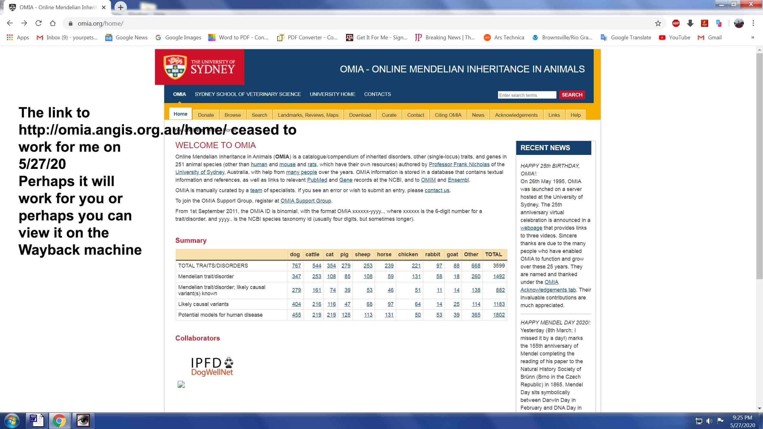