Common Hernias In Dogs And Cats
Umbilical, Inguinal, Perineal, Diaphragmatic And Pericardial-peritoneal Hernias And What Needs To Be Done
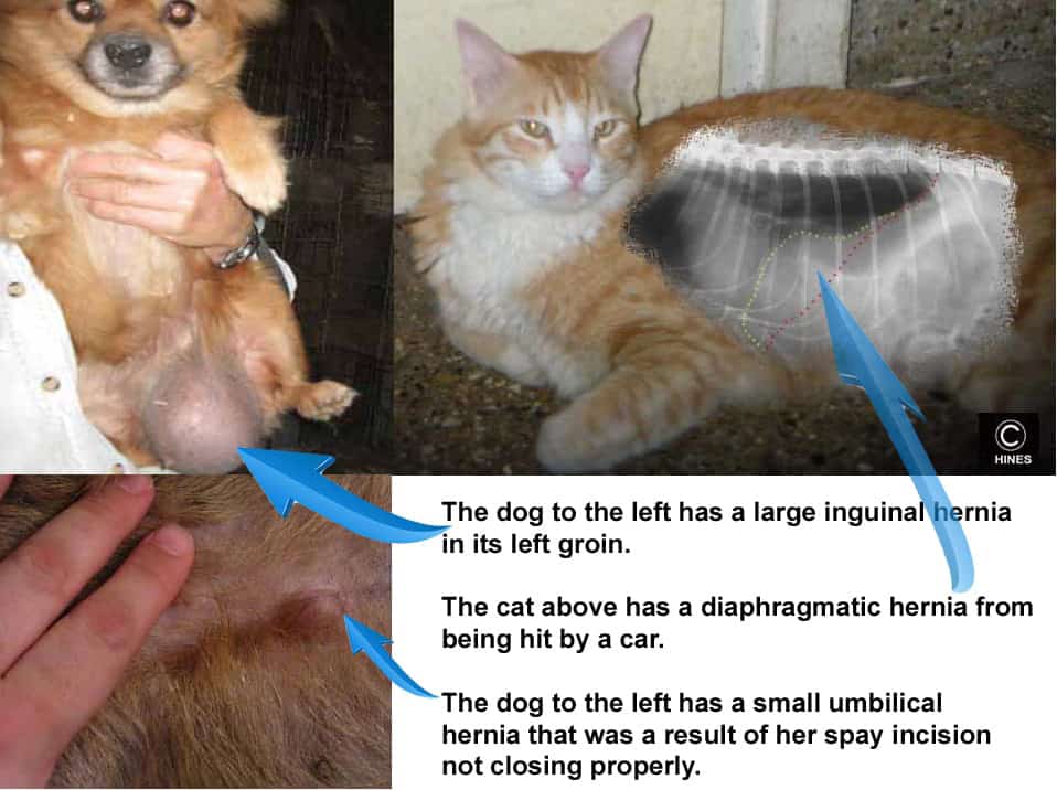
Ron Hines DVM PhD
What Are Some Common Hernias In Dogs And Cats?
Hernias are bulges and tears in your pet’s body wall or organs that allow tissue to pass into areas where they do not belong.
Hernias are a bit similar to sidewall bulges on an automobile tire. In both situations, a supportive barrier has been damage enough to lose its ability to hold back contents. Some hernias are minor inconveniences, while others can be life-threatening. Some hernias are present from birth (congenital hernias) while others are the result of injury or perhaps a surgical incision that failed to heal completely. When the hernia’s contents can be pressed back into its normal position it is called a reducible hernia. With time, hernias often form a tough fibrous ring around their edge. In those situations, the contents of the hernia sac sometimes become “trapped” in the pocket or sac and can no longer be manually manipulated to where they belong (a non-reducible hernia). If the tissue trapped inside the hernias does not receive an adequate blood supply, that situation is called a strangulated hernias. Strangulated hernias are an emergency situation. They also occur when organs such as the bladder or intestine “flip over” or twist within the hernia space and can no longer empty.
Umbilical Hernias:
The umbilicus, or naval is your dog or cat’s belly button; it’s source of nutrients and oxygen and exit for metabolic waste while still in the womb. Congenital umbilical hernias are the most common of all the hernias that veterinarians encounter. Since this problem might be an inherited trait, many feel that it is wise not to breed pets that are born with this condition. Very few cat or dog breeders take that advice.
Dogs and cats with umbilical hernias have a soft, painless swelling – a bulge or bubble over the point where their belly scar should be. The swelling may come up and go down depending on your pet’s position and how much it has recently eaten. You might be able to push it back in gently with your finger.
Small umbilical hernias contain nothing but a portion of the fatty veil we all have in our abdomens called an omentum. The fatty omentum normally drapes over the intestines like a veil. When pets become obese, their omentum fills with excess fat and enlarges. So, you might notice your dog or your cat’s umbilical hernia enlarge if your pet should become chubby.
Many small umbilical hernias are not a serious problem. They sometimes close by themselves as your young pet matures. Sometimes when they close, a tiny bleb of omental fat remains just below the skin. That is no cause for alarm. Many veterinarians repair these hernias when male dogs and cats are about 18 weeks of age. By then, they are old enough to make the general anesthesia required for this surgery safe. In female pets I often spay them right through the hernia defect and repair the hernia on my way out. I spay dogs at 8-12 months of age. But in-and-out female cats I spay younger because they can become pregnant when they reach about 5 pounds. Veterinarians differ in their preferred timing for umbilical hernia repair. There is a common misconception that cutting the umbilical cord off too close to the pet’s body is the cause of this condition. Since genetics plays a part in some umbilical hernias, veterinarians tend to encounter them more in purebred dogs and cats than in pets that resulted from accidental cross or random breeding.
As I mentioned earlier, larger umbilical hernias can strangulate important body organs. That is, cut off their supply of vital oxygen, blood and nutrients. That might occur when a loop of the pet’s intestines or portion of another body organ, got pinched off or twisted within the hernia sac. In those cases, the hernia’s fibrous ring can squeeze off the blood supply to the strangulated segment of the pet’s intestine, causing cell death and necrosis. As that damaged tissue within the sac swells, edema fluid squeezed it even tighter. That rarely occurs, but if it does, it is a life-threatening emergency. If you notice such a situation when your regular veterinary clinic is not open, it can’t wait until morning.
Large body wall hernias, with the skin intact, are frightening. But they can pose less immediate danger than the medium size ones. The large ones put no pressure on the intestines and portions of other body organs that may be inside their cavity. However, the trauma that caused them probably did cause substantial damage to internal organs that your veterinarian will deal with first. Large hernias can be a challenge for your vet to close because of a scarcity of available subcutaneous tissue to lap over the defect. When hernias do not have sufficient body wall tissue to overlap and sew together soundly, synthetic fabric webbing is used in dogs and cats – just as it is in hernia repair in humans.
Inguinal And Femoral Hernias:
Hernias in the groin commonly occur in female dogs that are, or have, been pregnant with multiple large litters, or dogs that experienced bloat or constipation. They can occur in cats as well. Veterinarians occasionally see the problem in males. In males, they are usually genetically based. In all cases, tissue that belongs in the rear of your pet’s abdominal cavity protrudes out through a weaken area surrounding your pet’s femoral artery and nerve. Usually, the hernia sac contains nothing but fat. It is usually reducible back into the abdomen with gentle finger pressure when your pet is on its back. Under general anesthesia, this sac can be very carefully dissected out with scissors and scalpel until it resembles a small balloon attached to the inner thigh. Then it can be carefully replaced into the abdomen. One has to be very cautious when darning the remaining hole shut, not to pinch the femoral artery or nerve. It is quite common for a second hernia to form later in the unaffected, opposite groin. So, to be safe, I check both sides very carefully and if any weakness exists on the opposite leg, I reinforced that area too (non-absorbable “purse string” sutures). Veterinarians occasionally see inguinal hernias in immature pekingeses and other small breeds – too young to be due to the increased abdominal pressure of pregnancy or abdominal distention. These hernias also occur occasionally in males. When they do, the surgery is the same. Post-surgical scaring in your pet should reinforce and block future hernias at the site. They should not reoccur. These hernias must be repaired very delicately by your veterinarian so as not to restrict blood flow and nerve impulses to and from the leg. As I mentioned, the surgery is especially challenging in toy dogs and cats. (read as it occurs in human here)
Perineal Hernias:
Perineal hernias occur just lateral to your pet’s anus. Many more are seen in elderly dogs than in cats. They are most common in male dogs that have not been castrated. In those pets, they often occur secondarily to an enlarged prostate – although straining to defecate due to constipation is another cause. Less commonly, veterinarians see perineal hernias in female dogs as well. Others believe that poorly developed muscle mass in the rump area and male hormones predispose to this condition. Perineal hernias can be confused with enlarged, infected or ruptured anal sacs (glands) or the anal sac tumors that sometimes form there. Inherited weakness in the structures that form the ligamentous ring around the anus are also thought to contribute to perineal hernias. Sometimes only one side is affected but, more commonly, both sides eventually prolapse (e.g. bulge) to some degree. As I mentioned, these hernias sometimes occurs when your pet excessively strains to pass hard stools or when a chronically inflamed anal sac causes constant straining. Sometimes the straining (tenesmus) could be due to a general inflammation of the anal region. Those pets most often scoot. That could also be due to feeding an inappropriate diet.
It is usually just abdominal fat that works its way into your pet’s perineal hernia sac initially; but there are cases in which the bladder or portions of its intestine eventually enter the sac as well. When that occurs, the problem becomes a medical emergency because your pet cannot urinate or defecate. Unless there is substantial swelling, the contents of the sac can be slid back into the abdomen manually when the pet’s rear end is elevated. Do not attempt that at home, it is the job of your veterinarian.
The technique for repair of these hernias is similar to that for femoral hernias. But in these cases, the difficult part is finding enough tissue surrounding the anus to unite with the internal pelvic structures. It is a difficult, tedious, tunneling operation because the pelvic bones prevent good exposure of the surgical area. Many veterinarians use non-absorbable suture that lasts your pet’s lifetime to “darn” these defects closed. One must be very careful not to injure the nerves of the rectum and anus during this surgery or your pet might become fecally incontinent. The maintenance of complete sterility during is operation is next to impossible, since this is a highly contaminated area. So pre and post surgical antibiotics are generally prescribed. Sometimes two or more operations are required before the defect can be completely closed. When one surgically correct this condition it is common to remove the anal sacs as well since they might have been the original cause of straining. As I mentioned, improper diet is the root of many of these cases. So be sure your pet get plenty of vegetable fiber in its diet and stays well hydrated. (read here)
Diaphragmatic Hernias:
Although hernias of the diaphragm can be congenital (a birth defect), all of them that I have repaired occurred subsequent to a car accident or a fall off the balcony. These diaphragmatic tears occur when pressure within the pet’s abdomen suddenly rises, pressing the organs of the abdomen forcefully against the diaphragm and rupturing it. These can be the most difficult of all hernias to repair. Pets with this condition often arrive at their animal hospital panting or gasping for breath. They are reluctant to lie down, because on their side, they have even more difficulty obtaining sufficient oxygen. Often, they become quite agitated if their rear legs are elevated like a wheelbarrow. In fact, that is a simple test veterinarians often utilize when they are suspicious that a diaphragmatic tear has occurred. X-rays confirm the diagnosis when chest organs are pressed forward and evidence of the stomach and/or other abdominal organs can be seen in the posterior chest cavity. Those x-rays also usually show indistinct areas of the diaphragm at the point of the tear.
In the diagram I made of the cat at the top of this page, everything between the red and the yellow dotted lines belongs behind the red dotted line (the diaphragm). In that cat, the tear in the diaphragm has allowed organs from the abdomen to press against the heart and squeeze the cat’s lungs so that they cannot fully inflate. If the x-rays are not as obvious as this one, veterinarians often repeat them after giving the pet an oral dose of barium sulfate. On subsequent x-rays, the barium will outline the intestinal tract and show your veterinarian if segment(s) of the pet’s intestine have passed through the tear and into your pet’s chest cavity. These tears can be of any size. They most commonly occur where the diaphragm attaches to the rib cage – a weak point. When corrective surgery is performed, one assistant must “bag” the pet (breath for the animal). The surgical approach can be quite difficult because the liver and stomach tend to block access to the region of the tear. But once a tear has been repaired your pet can resume a normal life.
Scrotal Hernias:
Scrotal hernias are much more common in horses and humans than in dogs or cats. I have never encountered one in a dog or a cat, but I am told that they do occasionally occur. Repair of a scrotal hernia would be similar to repair of an inguinal or femoral hernia. Again, such a hernia would only become life-threatening if a loop of intestine or the bladder passed down into the scrotum and became constricted. If a dog or cat with a scrotal hernia was presented to me, I would just suggest that the dog or cat be neutered through the same surgical incision. Why perpetuate what was likely a birth defect.
Pericardial-peritoneal Hernias:
Most pericardia-peritoneal hernias occur as a congenital defect (a problem your pet was born with). In this particular problem, an inherited pathway or tunnel runs from your pet’s abdomen all the way to the sac that surrounds its heart (the pericardial sac). Symptoms of the disease are similar to the symptoms as one might see in heart or lung failure, because the intestines/and/or liver lobe(s) that have migrated into your pet’s chest surround and press on its heart and lungs. Repair of such a defect would best be attempted at a large central veterinary specialty practice or university veterinary school.
You are on the Vetspace animal health website
Visiting the products that you see displayed on this website help pay the cost of keeping these articles on the Internet.